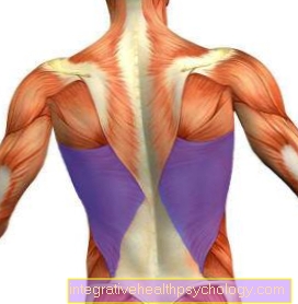
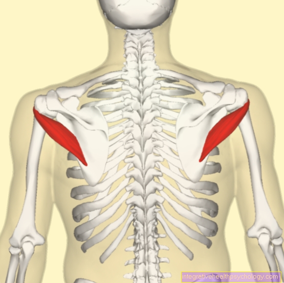
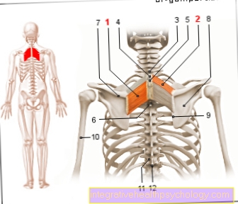
Category : Anatomy-Lexicon

M. teres major

M. teres major

The large round muscle (M. teres major) is one of the rear shoulder muscles. It connects the humerus with the lower part of the shoulder blade and, when contracting, raises the upper arm behind the body or pulls the raised arm back to the K

Collagenase

Collagenase

A collagenase is an enzyme that is able to break down collagen. It breaks bonds between two amino acids. One of these amino acids is always proline, while the second can vary.

The nucleus

The nucleus

The cell nucleus or nucleus is the largest organelle in a cell and is located in the middle of the cell plasma. It is surrounded by two shells and contains the cell's genetic information. This is important for making proteins for the body.

Rhomboid muscle

Rhomboid muscle

The rhomboid muscle (M. rhomboideus major and minor) runs from the spinous processes of the cervical and thoracic vertebrae to the inner edge of the shoulder blades. It is completely supported by the M.trapezius and its main function is to stabilize the shoulder blade

External rotation

External rotation

External rotation is the rotation of a body part around its own axis and away from the body. The external rotation is the opposite movement to the internal rotation and can be carried out by means of ball joints. For example, when tightening the shoulders

Hand nerves

Hand nerves

The hand nerves are responsible for the sensitive and motorized supply of the hand and arise from the arm nerve plexus (brachial plexus). A distinction is made between the median, radial and ulnar nerves. Damage usually leads to a loss of function

Cervical vertebrae

Cervical vertebrae

The cervical spine is part of the spine and consists of seven cervical vertebrae. The first cervical vertebrae are called the Atlas and Axis and are important for head movements and support the entire head. Often, accidents damage the cervical vertebrae, which is often the case

Radial nerve

Radial nerve

The radial nerve is an important arm nerve that transmits information from the skin to the brain and is important for muscle movement. In the event of damage, the "hand drop" occurs, in which the elbow joint is stretched

Lumbar spine (lumbar spine) x-ray with scoliosis

Lumbar spine (lumbar spine) x-ray with scoliosis

the medical information portal. X-rays on the subject of scoliosis of the lumbar spine.

Broad back muscle

Broad back muscle

The broad back muscle (M. latissimus dorsi) is a large, superficial back muscle that originates in the spine and attaches to the upper arm and sides of the ribs. Its main function is the adduction of the raised arm as well as the retroversion

Trapezius

Trapezius

the sports medicine information portal. A lot of helpful information on the topic of cap muscles explained in an understandable way.

Cruciate ligament

Cruciate ligament

Humans have two cruciate ligaments in each knee joint, an anterior and a posterior cruciate ligament. They stabilize the knee joint and prevent excessive inward rotation. Cruciate ligament tears are one of the most common knee ligament injuries, although mostly

Disc meniscus

Disc meniscus

A disc meniscus is a standard variant of the meniscus. Usually the menisci are shaped like a crescent moon. The disc meniscus, on the other hand, has a disc shape and is larger. Usually it does not cause pain and will

Quadriceps

Quadriceps

The quadriceps represent the largest and most powerful muscle in our body. The quadriceps is the most well-trained muscle in leg muscle training. The following are the functions, innervation, stretching and proper training of the quadriceps

Inner band knee

Inner band knee

The inner ligament of the knee joint connects the thigh with the shin. It is fused with the joint capsule and taut in the extended knee together with the outer ligament. In many sports the inner band is due to strong pressure

Outer ligament of the knee

Outer ligament of the knee

The outer ligaments of the two knee joints connect the thigh to the fibula. Together, the inner and outer ligaments are also called "collateral ligaments". They provide lateral stability to the knee joint when it is extended and limit rotation

Kneecap jumped out

Kneecap jumped out

The doctor describes the condition of a popped kneecap as a dislocation of the kneecap or patella or a dislocation of the kneecap or patella. Such a dislocation of the kneecap is relatively common and has two basic mechanisms

Taping the ankle

Taping the ankle

The ankle is one of the most commonly injured joints. These include bruises, slight stretching of the ligaments or ligament tears and injuries to the joint capsule. Kinesio tapes gently massage the affected areas

Broken fibula

Broken fibula

In medicine, a fracture of the fibula is called a fibular fracture. Most often it occurs in connection with a fracture of the upper ankle joint. In the case of serious injuries, the connective tissue membrane between the shinbone can also be affected

Rear foot

Rear foot

Anatomically, the rear foot corresponds to part of the tarsus. The term hindfoot comes from everyday clinical practice. Here the term two of the tarsal bones are described, namely the talus (ankle bone), the part of the ankle joint