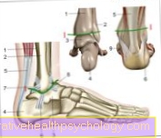
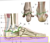
Figure ankle
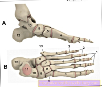
- Toe phalanx -
Phalanx distalis - Middle toe -
Phalanx media - Phalanx -
Phal. proximalis
(1st - 3rd toe bones -
Phalanges) - Metatarsal bones -
Os metatarsi - Inner sphenoid bone -
Medial cuneiform bone - Middle sphenoid bone -
Os cuneiform intermedium - External sphenoid bone -
Os cuneiform laterale - Cuboid bone - Os cuboideum
- Scaphoid bone - Navicular bone
- Ankle bone - Talus
- Ankle roll - Trochlea tali
- Heel bone - Calcaneus
- Protrusion on the 5th metatarsal -
Tuberositas ossis metatarsalis quinti (V)
You can find an overview of all Dr-Gumpert images at: medical illustrations
Related images

Illustration
Achilles tendon

Illustration
Torn ligament

Illustration
Heel spur

Illustration
Foot pain
Illustration
Upper ankle

Illustration
Instep pain
Illustration
Lower ankle