Anatomy and function of the human skin
General information about the skin
The skin of the human body has a total area of 1.5 to 2 m2. The total weight is around 3.5 to 10 kg.
The surface shows an individually different relief. This relief is genetically determined. The skin is divided into two different types. On the one hand the hairless one Inguinal skinwhich is located on the palms of the hands and feet.
Here there is a so-called papillary ridge course, which divides the inguinal skin. This creates fingerprints that are genetically determined and individual for each person.
The rest of the skin surface is divided into irregular fields by furrows. In these furrows the so-called Field skin are the hairs.

Likewise, the skin is sensitive through annoy (Sensory nerves) divided into so-called dermatomes. Under Dermatome one understands the segmental skin area innervated (supplied) by a spinal nerve. Spinal nerves step out of the Spinal cord and run into their coverage area. Each spinal nerve consists of many afferent (leading away) Nerve fibers that reach the skin via various peripheral nerves.
Structure of the skin
The skin is made up of several layers made up of different tissues. On average, our skin thickness is 1.5 to 4 mm.
The skin is roughly divided from the outside inwards into the upper skin (epidermis), dermis (dermis) and the subcutaneous tissue.
Epidermis
The epidermis, in turn, is divided into four to five layers, depending on the type of cells that can be found in these layers.
From the outside in, these are: horny layer, glossy layer, granular cell layer, prickly cell layer and basal layer.
The horny layer, which is too extreme on our skin, consists mainly of dead cells. This layer is particularly pronounced in the cornea, which we find on the soles of our feet, for example, because the skin there is exposed to particular stress. The dead cells peel off our skin over time, but are constantly renewed from below by new cells, which are created by cell division in the basal layer.
In the basal layer there are also pigment-forming cells, the so-called "melanocytes", which give our skin its color.
The glossy layer is only in the so-called groin skin, which can be found on the palms of the hands and soles of the feet. In contrast, the skin in all other regions of our body is called field skin. It covers about 96% of our body surface.
In the epidermis, pain signals and light touches that hit the skin from outside are picked up and passed on to the brain.
Dermis
The dermis consists mainly of connective tissue fibers and is responsible for anchoring the epidermis.
The blood vessels, which are essential for the nutrition of our epidermis, run in this layer. It is also important for regulating the temperature of the skin. Hair roots, sebum glands and sweat glands are embedded in the dermis.
In addition, sensations of touch and pressure are recorded in this layer and transmitted to our brain.
The dermis is divided into a papilla layer and a mesh layer.
The papillary layer contains so-called papillary bodies, which are arranged in longitudinal rows in the groin skin on the palm of the hand and the sole of the foot and can thus be seen there as "skin ridges". Our fingerprint is created on the basis of these "skin ridges".
Subcutis (subcutaneous tissue)
The subcutaneous tissue is mainly made up of subcutaneous fat and loose connective tissue. Nerves and larger blood vessels run in it to supply the layers above. Similar to the dermis, sensory cells can be found here, which, however, tend to absorb and transmit strong pressure sensations.
Dermatomes
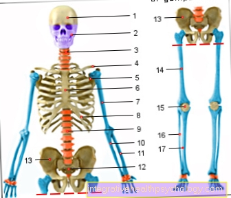
The Dermatomes map the sensory area of individual spinal nerves.
A sensory area is understood to be the supply area of a nerve with feeling.
This is clearly illustrated in the adjacent picture.
Red is the supply area of the nerves Cervical spine, blue the area of the Thoracic spine.
A failure / damage leads to a sensory disorder of the skin precisely in the area of the respective nerve depicted.
Read a lot more information on this topic at: Dermatome
Figure skin
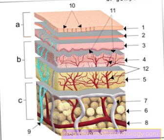
a - epidermis (1st - 3rd) - epidermis
b - dermis (4th - 5th) - Dermis
c - subcutaneous tissue (6.) - Tela subcutanea
- Horny layer - Stratum corneum
- Cornifying layer
(light layer
and granular layer) -
Stratum lucidum and
Stratum granulosum - Germ layer (prickly cell layer
and base layer) -
Stratum spinosum and
Stratum basale - Papillary layer -
Stratum papillary - Network layer - Stratum reticularre
- Subcutaneous tissue - Tela subcutanea
- Lymph vessel - Vas lymphaticum
- Artery - Artery
- Cutaneous nerve - Cutaneous nerve
- Duct of a sweat gland -
Ductus sudorifer - Papillae of the dermis -
Papillae (dermidis) - Vascular network of the dermis -
Subpapillary venous plexus
You can find an overview of all Dr-Gumpert images at: medical illustrations
Composition of the skin
Our skin consists of two layers:
-
the epidermis, the upper layer, epidermis
-
the dermis, the lower layer, dermis
These each consist of further thin layers. Further below is the subcutaneous fatty tissue.
1. The epidermis
Structure and cells
The epidermis, also called the epidermis, is a multi-layer structure that has the ability to keratinize.
It consists of five different histologically (under the microscope) visible cell layers. The epidermis is of different thickness in different parts of the body.
In places that are subject to a lot of stress (hands, feet) it is thicker, in places that are less stressed (arms, face) it is rather thin. The thickness varies from 30 to 300 micrometers. As a so-called proliferation tissue (proliferation means increase) it is subject to constant renewal. There are many nerves in the epidermis, but no blood vessels. The supply takes place through diffusion (passive transport) from the layer below, the dermis.
The different layers of the epidermis also contain different types of cells. The main ingredient, however, represent the Keratinocytes (Horn cells). These cells migrate through the epidermis to the surface of the skin, changing their structure. When they reach the surface, they are peeled off as horn scales.
The name of the cells (keratinocytes) during the migration correlates with the layer in which they are located:
- Basal cell (regeneration layer)
- Prickly cell (prickle layer)
- Granule cell (granular layer)
- Horny cell (horny layer)
The duration of such a hike is usually about 5 to 7 weeks. The keratinocytes are anchored to the dermis by hemidesmosomes on the basement membrane. This is how their hold is secured.
Another component of the skin are among others the Melanocytes. These large, bright cells contain melanosomes, in which melanin is synthesized and stored.
Melanin is the skin pigment that gives the skin its actual brown color.
The melanin is then given to the neighboring keratinocytes. Melanin is a pigment that, for example, causes the skin to tan.
Also Langerhans cells are located in the epidermis. They play an essential role in allergies. For those particularly interested: The Langerhans cells are responsible for type IV allergies (e.g. allergic contact eczema).
T lymphocytes have an immunological function and occur occasionally in the epidermis, but mainly in the dermis. They cooperate with the Langerhans cells.
Merkel cells are found in the innermost layer of the epidermis. They convey the tactile sensation.
Connection zone between dermis and epidermis
The two layers of the Skin (cutis) are closely related. Among other things, so-called Retel strips ensure this connection.
A Basement membrane (thin separating layer) between the layers controls the exchange of cells and molecules. It consists of 2 layers. One of these layers is connected to the next layer of skin with the help of anchoring filaments. The inner layer is with the Dermis and the outer layer with the one on the outside epidermis connected.
2. The dermis
The second part of the cutis (skin), the dermis, also called the dermis, is the connective tissue under the epidermis and extends in depth down to the subcutaneous fat (subcutaneous = under the cutis / skin). The main components are cells and connective tissue fibers, which are embedded in a gelatinous basic substance.
These are collagen fibers, elastic fibers and reticulin fibers. This ensures the tear resistance and reversible (restorable) deformability of the skin.
The dermis is divided into two layers:
- The papillary layer (stratum papillare), which rests against the epidermis and
- the braided layer (stratum reticulare), which is directly adjacent to the subcutaneous tissue. Hair follicles and sweat glands arise in the braided layer.
There are also plexuses of vessels (vascular plexus) in the dermis. They serve to supply the skin with nutrients and to regulate temperature.
Subcutis - subcutaneous tissue
This so-called subcutaneous tissue connects to the stratum reticulare of the dermis. It consists of loose connective and subcutaneous fat tissue.
Functions of the skin
The skin has a wide variety of functions, which can be explained by the individual components in the various layers.
Due to its natural skin flora and its somewhat acidic pH value, it represents a protective barrier against bacteria, for example. The skin contains cells of the immune system and is therefore part of our immune system.
The horny layer protects us from dehydration and injuries. Sweat glands are important to prevent overheating and sebum glands oil our skin.
Read more about the topic here: Anatomy and function of the skin glands
Not only the sweat glands are decisive for temperature regulation, but also the subcutaneous fatty tissue and the blood vessels, which run close to the surface and can regulate the heat emission through the blood circulation.
The hair and many sensory cells in different layers create contact with the outside world, which gives us the opportunity to absorb various stimuli, such as pain, touch, pressure and temperature sensation.
Furthermore, our skin protects us from UV rays. When exposed to the sun, it reacts with tanning, as UV rays would otherwise damage our skin very quickly.
In addition, the skin basically envelops our entire body from the outside, so that it represents a barrier to the environment. The skin can withstand some mechanical stress, but it cannot withstand blunt or sharp force. It then comes to wounds, such as Bruise wounds, stab wounds or a laceration.
Skin in balance - what does that mean?

There are so-called skin appendages in the layer of the epidermis. These include, for example, glands, which secrete fatty substances, and hair follicles.
The epidermis, with its horny layer, the secreted fat and its acidic pH value, serves as protection against external influences.
The exact pH value is now somewhat controversial. For a long time it was assumed that it was between 5 and 6, but there are now studies that suggest a pH value below 5.
In any case, it is in the acidic range and on the one hand has a protective function against certain pathogens, on the other hand it allows "desired" bacteria that belong to the normal skin flora to survive.
Another vital function of the epidermis is to protect it from dehydration.
Without the top layer of skin, up to 20 liters of water would be lost through the body surface every day. This explains why people with burns are at high risk of becoming dehydrated (to dry out) and therefore have to be supplied with a lot of water.
The dermis lies beneath the epidermis. It mainly contains fibroblasts, cells that produce connective tissue, especially collagen. But cells of the immune system, so-called histiocysts and mast cells, also develop here. The dermis also contains nerves and blood vessels.
As already mentioned, the skin has important functions in the area of homeostasis. It has a large part in regulating body temperature. In particular, it intervenes in a regulating manner through evaporation of water.
The skin is also extremely important for the absorption of stimuli. Whether touch, pain or temperature. This is done through receptor cells.
The skin is densely populated with microorganisms.
This sounds dangerous at first, but it isn't. This is known as normal skin flora. The bacteria that belong to this normal flora are not harmful. They are known as commensals. This means that they benefit from the fact that they colonize the human skin, but neither use nor harm the human being.
In part, they have a protective influence by protecting against the penetration of pathogenic germs.
The skin has a multitude of functions (please refer: Functions of the skin) that can only be guaranteed if it is in balance. The pH value has to be right, the surface of the skin has to be intact and the normal, resident flora of the skin also plays a role in a balanced complexion.
Symptoms and disorders of the skin
The skin cancer
There are different types of skin cancer, which are classified based on the cells it originates from. A distinction must be made between benign and malignant (malignant) types of cancer.
The most common skin cancer is basalioma, which occurs as a result of uncontrolled cell division in the basal cell layer. The basalioma is only partially malignant as it can infiltrate the surrounding tissue, but only in the rarest of cases does it form metastases.
Basalioma usually develops in areas that are heavily exposed to the sun and thus UV rays, such as the facial region.
On the other hand, there is malignant melanoma, which is a very malignant tumor of the melanocytes (pigment cells). It grows infiltratively and metastasizes prematurely.
As with all types of cancer, the early detection of possible degenerations is important. It is therefore advisable to pay attention to skin changes and to go to the dermatologist if anything is abnormal.
Harmless pigment spots can be distinguished from suspicious pigment marks by: regular, symmetrical shape and sharp, clear edges, as well as uniform coloring and no change in size, color, shape or thickness.
Read more about the topic here: Detect and treat skin cancer early on
Skin itches
Itching (Pruritus) is an unpleasant sensory perception that would like to be answered with mechanical resistance in the sense of scratching.
It was originally used to remove foreign bodies or parasites.
However, there is also chronic itching that lasts for at least six months and is no longer triggered by an adequate stimulus.
The nerve fibers used to detect itching are among the pain receptors (Nociceptors) and are mainly located within the top two skin layers, the epidermis and the dermis. The stimuli are absorbed via unmarked C-fibers and passed on to the central nervous system where there are pruritus-specific areas.
There are numerous hormonal triggers that can cause itching. The best known is probably histamine. That is why antihistamines are often prescribed to treat itching, i.e. drugs that work against the histamine.
But since numerous other substances, such as serotonin, adrenaline, prostaglandins and dopamine, can initiate itching, these drugs are often not effective.
A variety of conditions can cause itching. Those that are localized in the skin area, i.e. dermatological diseases, but also internal and psychiatric diseases.
As an example, some diseases are listed here that can be accompanied by itching: The dermatological diseases that often show itching as a symptom include the drug eruption (Medication rash), Neurodermatitis (atopic eczema), Hives (Urticaria), Psoriasis (psoriasis) and scabies (Scabies).
Internal diseases that can be accompanied by itching include kidney failure, liver diseases such as primary biliary cirrhosis, malignant diseases such as leukemia and Hodgkin's disease, metabolic diseases such as diabetes mellitus and iron deficiency.
The psychiatric illnesses that can be associated with itching include schizophrenia, depression and anorexia, among others.
Numerous medications can also cause itching. For example ACE inhibitors, antibiotics, calcium antagonists, beta blockers, antimycotics, immunomodulators, lipid lowering agents, psychotropic drugs and many others.
In dermatological diseases, the itching is often more localized, i.e. particularly pronounced in certain areas, while in internal diseases it usually affects the whole body.
The therapy of the itching depends mainly on the cause. The respective disease that leads to itching must be treated specifically. This is known as causal therapy.
Purely symptomatic therapy aims to relieve the itching, but does not get the cause out of the way. Various creams are available for symptomatic therapy: There are creams that have a slightly numbing effect (contain lidocaine), those that contain anti-inflammatory glucocorticoids such as cortisone, or those that have immunomodulators such as tacrolimus as an active ingredient.
Furthermore, as mentioned above, antihistamines such as cetirizine can provide a remedy; these are usually administered in tablet form. Psychotropic drugs such as neuroleptics or tricyclic antidepressants can also help. All in all, if itching is a symptom, it is always necessary to look for the causal disease and, if possible, treat it causally in order to treat the itching in the long term.
Read more on this topic at: Skin itches
Skin burns
The skin is in constant contact with the environment and is therefore exposed to many stimuli.
Burning skin is a sign that the skin has come into contact with a substance that it cannot tolerate. These can be intolerance reactions or allergic reactions, for example to food or substances in care products or cosmetics.
Such a hypersensitivity reaction usually manifests itself in skin changes that redden the skin or develop blisters.
Find out more about the topic here: Blistered rash
Burning skin can also occur with the second disease or late sequela of chickenpox, known as "shingles". Those who suffered from chickenpox in their childhood are immune to chickenpox again, but the virus remains in the body for a lifetime.
If the immune system is weakened, for example due to stress or a cold, the virus can be responsible for the occurrence of shingles. It manifests itself in a belt-shaped rash with reddish blisters, usually in the abdominal region, which burns and itches a lot.
Another possibility of skin burning sensation can be due to hypersensitivity of nerves. In this case, the burning sensation is often accompanied by a tingling sensation and / or numbness. In the event of abnormalities such as severe burning or rashes, a doctor should be consulted and the causes clarified.
Read more about the topic here: Skin burns
The skin fungus
Fungi that are pathogenic to humans, i.e. those that are relevant to damage to human health, are divided into three classes:
- Dermatophytes
- Yeasts
- Molds.
Most fungi are facultative pathogenic, which means that they cannot infect a healthy person, but can make a person with a weakened immune system or a disturbed skin defenses sick.
Dermatophytes only attack the skin, hair and nails while yeasts such as Candida albicans and molds such as Aspergillus flavus can also attack internal organs.
Skin fungus is mainly caused by dermatophytes and is then referred to as tinea. The most common causative agent of tinea in Central Europe is the fungus Trichopyhton rubrum.
The fungal attack on the skin can be classified according to the penetration depth of the pathogen. A distinction is made here between superficial tinea (Tinea superficialis) and deep ringworm (Tinea profunda).
Tinea superficialias often has an almost round, reddish-brown foci on the skin that has a pronounced edge. However, there are numerous other manifestations of the superficial skin fungus.
The more invasive form of tinea is called tinea profunda (deep down), the pathogens penetrate deeper into the skin. It is mainly found on more hairy parts of the body such as the beard or scalp.
In addition, the skin fungus can be divided according to location. The most common place for fungal infections is in the spaces between the toes (Interdigital spaces).
A fungus found in this area is called tinea pedis (Athlete's foot) designated. The athlete's foot can be dangerous insofar as it can create entry points for bacterial pathogens. This can lead to bacterial superinfections that spread in the body. A typical example of a disease whose pathogens often enter the body through such an entry port is erysipelas.
Furthermore, after localization, tinea palmoplantaris, which is accompanied by scaling on the soles of the hands and feet, tinea capitis, which is noticeable by approximately round hairless foci on the scalp, tinea corporis, which is often round reddish foci on the trunk and arms and Legs becomes noticeable and tinea ungium of the toenails (Nail mycosis) can be distinguished.
A smear from the edge of the affected skin area with a subsequent microscopic examination can be used to determine whether the skin is infected with fungal infections.
In uncomplicated cases, local (topical) treated, i.e. not with tablets but, for example, with solutions or creams. It depends on which pathogen it is, because yeasts (Candida) can cause skin infections and some respond to a different therapy than the dermatophytes just discussed.
However, broad-spectrum antifungal agents that work against both types of fungus are now widely used. These include ciclopiroxamine, clotrimazole as well as terbinafine and amorolfine. Fluconazole is particularly suitable for treating yeast infections.
They are available - depending on the preparation - as a cream, solution or nail polish. However, some types of skin fungus can only be treated systemically, i.e. using tablets, with the duration of therapy usually extending over several weeks. It is usually combined with local therapy.
Read more on this topic at: Skin fungus
Bleach skin

Skin bleaching is also called Skin lightening designated. It mainly serves cosmetic purposes, but sometimes also occurs with morbid Overproduction of the dye Melanin (Hyperpigmentation) to use.
The history of the heap lightening is probably due to the fact that in earlier epochs a very light complexion than Ideal of beauty was true. The well-off people were often of a very fair complexion and the "workers" were mostly tanned by the sun. It was a bright one Skin color thus also a bit of a figurehead for social status.
Skin lighteners bring significantly more sales worldwide than skin tanning and sun protection products. Only one Active ingredient approved in Germany to lighten the skin Pigmanorm. It contains the active ingredients hydroquinone, hydrocortisone and tretionine and is used in melanin-related hyperpigmentation. It is only on in normal skin applied to small areas of the skin and should be dosed carefully and used for a limited time.
Numerous other means are not approved in many countries and are sometimes massive Side effects hand in hand. They contain, among other things toxic substances such as mercury, benzenes and hydrogen peroxide. A side effect that is common to almost all of these remedies is this significant inhibition of the skin's defense against UV radiation. This is because the lightening agents destroy the body's own melanin, which provides UV protection. Can follow Skin burns and - with a latency of years - the emergence of Skin cancer be. Perhaps the most famous celebrity example of excessive skin bleaching use was Michael Jackson.