Radial artery
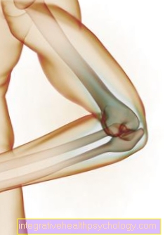
Anatomical course
It runs along the spoke (radius) on the front of the forearm under the Brachioradialis muscles. In its course it is supported by a superficial branch of the Radial nerve accompanied. It is in the foveola radialis ( Snuffbox) easy to feel. This is limited by the tendons of the extensor pollicis longus muscle and the extensor pollicis brevis muscle.
In its course the radial artery gives off the following branches:
- The Radial recurrent artery is a retrograde vessel and supplies the Elbow.
- Of the Ramus carpalis palmaris draws to the vascular network of the carpal bones and the
- Ramus palmaris superficialis pulls to the thumb.
- Of the Ramus carpalis dorsale supplies the outside of the hand.
- Another branch supplies the thumb (Arteria princeps pollicis) and the index finger (Radial artery indices). By the hand she goes into the palm of the hand (Arcus palmaris profundus), which lies on the base of the metacarpal bones and thus also supplies the palm of the hand with oxygenated blood.
- Some smaller branches that Arteriae metacarpales palmares run between the metacarpal bones and anastomose with the vascular branches of the superficial palm arch.
course
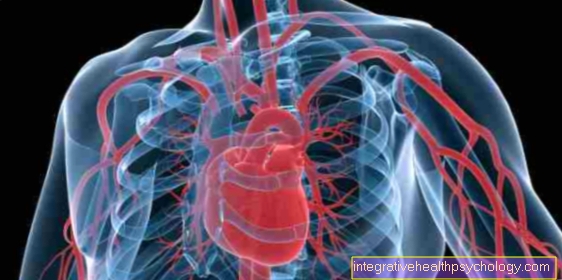
It splits in the crook of the elbow Brachial artery in two other arteries: The Radial artery and the A. ulnaris. The radial artery runs over the M. pronator teres and between the Flexor carpi radialis muscle and the M. brachioradialis. Of the M. brachioradialis is also the lead structure of the Radial artery. It runs first under the belly of the muscle and later next to its tendon. The Radial artery is accompanied by two veins that Vv. Radiales, and also lymphatic vessels. In the field of Snuffbox (Carpal) pulls the radial artery through the heads of the M. interosseus dorsalis I. and thereby succeeds at hand to palm. Here it gives off several branches, such as the A. princeps pollicis (for the thumb).
The Radial artery then forms together with the A. ulnaris the Arcus palmaris profundus that supplies the palm. Before the Extensor retinaculum (on the back of the wrist) the splits Ramus carpalis dorsalis from the Aa. metacarpales dorsales arise. These small arteries make up the thin ones Aa. digital dorsales and thereby supply the back of the fingers.
diameter
The radial artery is approximately in diameter 2 to 3 mm.
clinical importance
At the wrist can he Pulse The radial artery can be felt and measured well, so that here often arterial blood drawn for examination will, or a invasive blood pressure measurement can be created.
At dialysis- patients the arteria radialis is also usually used around a Cimino shunt to put on.
Palpate the pulse on the radial artery
The radial artery is a preferred location in medicine around the To feel the pulse. The easiest way to find the radial pulse is on the underside of the forearm, near the wrist. The radial artery runs along the forearm on the side of the Os radius (spoke), that is, on the same side as the thumb. The artery runs near the tendon of the Flexor carpi radialis muscle and des M. palmaris longus. When the wrist is flexed, the tendons of both muscles become particularly prominent.
In order to be able to feel the pulse, you have to use your fingers (preferably the index finger and middle finger, but never the thumb) on the thumb side directly next to the above-mentioned tendons, about one or two cross fingers below the ball of the thumb. The pulse can then be felt and counted on the fingertip. If the pulse disappears, you should reduce the pressure a little or put your fingers a little higher or lower.
Another place where you can feel the radial pulse is the Snuffbox. The tobacco box is located on the thumb side of the forearm below the thumb. This depression is particularly easy to find when the thumb is spread apart. As with the pulse measurement on the underside of the arm, you simply have to press your fingers lightly in this recess and then measure the pulse.
Removal of the radial artery (what, how does it work?)
The radial artery can be removed as part of a Bypass surgery be performed. The bypass operation is used to bridge a Constriction of the coronary arteries. If the coronary arteries no longer allow enough blood to pass through, there may be an undersupply of the heart muscles. To prevent this, an artery or vein is used as a bridge. For example, the artery is placed before and after the constriction so that the blood can continue to flow through this diversion.
More information can be found here: Coronary heart disease
The radial artery is removed minimally invasive, that means with just a few cuts. The surgeon can then endoscopically tie off and cut the arteries in two places. The two ends are closed to prevent bleeding. Various collaterals or anastomoses ensure that the supply areas of the radial artery continue to be supplied with blood (the A. ulnaris takes over the tasks of Radial artery). Due to anatomical variants in the supply, the blood flow via the ulnar artery should be tested.
The removal leaves a 2 to 3 cm long scar on the forearm. Another reason for the removal of the radial artery can be, for example, as part of the plastic / reconstructive surgery respectively. To restore missing parts of the skin, a piece of skin including the blood supply must be donated from another part of the body. In this context, the skin of the forearm with the radial artery can be considered.
You may also be interested in this article: Cardiac bypass - when is it used?
Radial artery and cardiac catheter
At a Cardiac catheterization it is an invasive (but not surgical) examination of the left or right heart with access via a vessel. For this you can use vessels in the groin or on the arm. A venous access is used to examine the right heart, and an arterial access is used to examine the left heart. A Left heart catheter examination can for example use the Radial artery respectively. Here a catheter is inserted into the radial artery and pushed to the heart under X-ray control. Once the catheter has reached the left heart, various examinations can be performed, such as one Pressure measurement of the left ventricle or a representation of the left ventricle using a contrast medium.
It is also possible to use this catheter to assess the coronary vessels with regard to coronary heart disease or to initiate interventional steps. For example, you can use a Balloon dilatation reopen and implant stents. At the end of the examination, the catheter is withdrawn and the wound closed. A pressure bandage is often applied to prevent rebleeding.