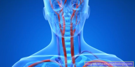
Vertebral artery
anatomy
The vertebral artery is one of the vessels that supply the brain with oxygenated blood from the heart. Their diameter is about 3-5mm. It is created in pairs, that is, there is a right and a left vertebral artery, which finally unite to form the basilar artery.
This vessel mainly supplies parts of the brain that lie in the posterior fossa. This includes areas of the cerebrum that are responsible, for example, for vision, such as the occipital lobe, or for hearing and speech understanding such as the temporal lobe. In addition, the cerebellum is also supplied by branches of the vertebral artery and the basilar artery. This is particularly important for balance and the coordination of movement sequences. The upper (cranial) parts of the brain stem, the bridge (pons) and the diencephalon (mensencephalon) are also supplied with blood from the basilar artery. These brain regions contain many cranial nerve nuclei that are responsible for the functions of the facial and eye muscles and the sensory organs in the face, as well as nerve pathways that interconnect the coordination of movement sequences.
Before it fuses to form the basilar artery, the vertebral artery also gives off branches to supply the upper spinal cord and parts of the brain stem, the medulla oblongata. This regulates basic and vital functions of the body such as breathing and circulatory regulation and the gag reflex.
course
The vertebral artery is a branch of the subclavian artery, which is also paired up. It rises approximately at the level of the depression between the collarbone (clavicle), neck muscles and cervical spine (supraclavicular fossa) and runs behind the anterior neck muscle (scalene muscle) to the cervical spine.
At the level of the 6th cervical vertebra, it enters an opening within this vertebra (foramen transversarium). All cervical vertebrae have this opening in their lateral process (processus transversus), which is why the arteria vertebralis can pull relatively protected along the cervical spine to the head through these superimposed holes. The superimposed holes are also known as the vertebral canal (Canalis vertebralis). On the head, the artery enters the posterior fossa through the foramen magnum at the transition from the neck to the head.
Sections
The vertebral artery is from the beginning in four segments (V1-V4).
Segment V1 describes the free course of the artery until it enters the intervertebral orifices. Changes to the inner vessel wall such as Calcifications as part of a arteriosclerosis on. It can also happen that the vascular wall loses its elasticity due to aging processes and kinks, which leads to a (functional) closure.
The V2 segment runs through the vertebral canal and can be narrowed here mainly due to age-related changes in the cervical vertebrae. The segments V2 and V3 (area of the first cervical vertebra where the arteria vertebralis loops around the first cervical vertebra) are most at risk for external injuries, for example in accidents, due to their anatomical proximity to the cervical spine.
The fourth segment is the section of the vertebral artery that runs inside the skull.
function
The vertebral artery supplies the brain and parts of the spinal cord with oxygen-rich blood. Mainly the cerebellum, brain stem and occipital lobes are supplied by the vertebral artery (see anatomy).
An important function of the vertebral artery is only relevant in the case of a certain clinical picture.
Does a patient suffer from the so-called Subclavian steal syndrome it ensures blood supply to the upper extremity by becoming part of a bypass circuit.
In this clinical picture, the artery from which the vertebral artery arises, the subclavian artery, is narrowed on one side or completely closed. Since the subclavian artery normally supplies the arm with blood, there is no blood there. In order to ensure the blood supply to the arm nonetheless, the vertebral artery is "tapped" on this side, which is why the syndrome is also referred to as the Vertebralisanzapf syndrome.
The blood flow in the vertebral artery is reversed and directs blood from the opposite vertebral artery into the poorly supplied arm. However, since the blood to supply the brain is lacking, it can lead to insufficient supply of the brain, especially with increased arm work, and the patients develop symptoms such as dizziness or headaches.
Vertebral artery syndrome
The so-called vertebral artery syndrome refers to a Circulatory disorder in this area. This can have various causes.
On the one hand, the poor blood circulation in one Vascular calcification (Arterial sclerosis), which narrow the diameter of the artery and make it harder for blood to flow. In this case one speaks of a vascular (vessel-related) arteria-vertebralis syndrome. On the other hand, the vertebral artery may be also constricted from the outside be, for example by a tumor, a Intervertebral disc of the Cervical spine or the Cervical vertebrae himself. Here one speaks of one Vertebral artery compression syndrome.
The symptoms of vertebral artery syndrome are similar to those of so-called basilar migraines, as the areas of the brain that are supplied with blood by the basilar artery are particularly affected. The main symptom is Attack-like dizzinesswhich is triggered by the reduced blood flow to the inner ear.
In the case of a compression syndrome, it is usually "vertebral dizziness". Age-related changes in the cervical vertebrae can lead to the formation of so-called osteophytes, bony protrusions that protrude into the spaces between the individual vertebrae and can compress the vertebral artery. Rotation of the head can increase this narrowing of the artery. Since, among other things, the inner ear with the organ of equilibrium is also supplied by the vertebral artery, this can trigger "vertebral dizziness".
There can also be many other non-specific symptoms such as a headache (especially on the back of the head), Visual disturbances, Ringing in the ears, nausea, Vomit, Unsteadiness (Ataxia) and Sensory disturbances occur. Approximately 50% of people with vertebralis syndrome also have one depressive mood in front.
If the doctor has made the suspected diagnosis of vertebral artery syndrome by means of a neurological examination, a Ultrasound examination and MRI examination searched for the cause. This then determines the therapy in the further course.
If it is a narrowing of the vertebral artery due to vascular calcification, it is often necessary to use a so-called Stent (Plastic tube) into the vessel. This will restore blood flow and symptoms will go away.
If the vertebral artery is constricted by a cervical vertebral body, conservative therapy without surgery is usually sufficient. The patient gets Painkiller such as Chiro and physiotherapy prescribed. Surgery is indicated if the cause of the vertebral artery compression syndrome is a severe herniated disc (prolapse) of the cervical spine or if there is a compressing tumor in the cervical spine.
Vertebral artery dissection
The dissection of an artery is called the Splitting of the inner vessel wall (Intima). This can lead to bleeding between the intima and media (middle vessel wall). This leads to a narrowing (stenosis) or in the worst case to complete closure of the vessel with circulatory disorders in the affected area of the brain.
The vertebral dissection affects primarily young adults and can occur spontaneously or, for example, in a car accident. The leading symptom of vertebral dissection are Headache in the back of the head. Besides, it can too Nausea, vomiting and dizziness come.
Usually used as therapy anticoagulants used, which have to be taken over a relatively long period of time (6-12 months depending on the agent). In rare cases, surgery and possibly the insertion of a stent into the vessel are necessary.
Clasp
Various mechanisms can lead to the occlusion of the vertebral artery. One mechanism, for example, is dissection, which has already been mentioned above.
Generally, infarcts (= vascular occlusions) in the area of the vertebral and basilar arteries are rare. They are usually the result of arteriosclerosis in other vessels. There, material from the vascular wall can be detached and washed into the vertebral artery as an embolus (vascular plug). The symptoms are similar to those of vertebral artery syndrome.