Arthroscopy
Synonyms
- reflection
- Knee reflection
- Shoulder mirroring
- Keyhole surgery
English: arthroscopy
definition
An arthroscope is a special endoscope. It consists of an optical system of rod lenses, a light source and usually a rinsing and suction device. In addition, the arthroscope has working channels through which surgical instruments can be inserted for minor surgical interventions. The optics of this endoscope are often connected to a monitor via a camera to make work easier. With this arthroscope, the doctor can look directly at the joint structures, similar to a camera.
Appointment with a knee specialist?
I would be happy to advise you!
Who am I?
My name is dr. Nicolas Gumpert. I am a specialist in orthopedics and the founder of .
Various television programs and print media report regularly about my work. On HR television you can see me every 6 weeks live on "Hallo Hessen".
But now enough is indicated ;-)
The knee joint is one of the joints with the greatest stress.
Therefore, the treatment of the knee joint (e.g. meniscus tear, cartilage damage, cruciate ligament damage, runner's knee, etc.) requires a lot of experience.
I treat a wide variety of knee diseases in a conservative way.
The aim of any treatment is treatment without surgery.
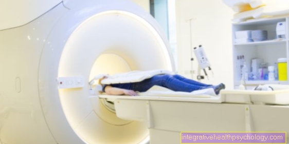
Which therapy achieves the best results in the long term can only be determined after looking at all of the information (Examination, X-ray, ultrasound, MRI, etc.) be assessed.
You can find me in:
- Lumedis - your orthopedic surgeon
Kaiserstrasse 14
60311 Frankfurt am Main
Directly to the online appointment arrangement
Unfortunately, it is currently only possible to make an appointment with private health insurers. I hope for your understanding!
Further information about myself can be found at Dr. Nicolas Gumpert
Arthroscopy
What is an arthroscopy?
An arthroscopy means the "Knee arthroscopy", i.e. a look inside the Knees with the help of an optic.
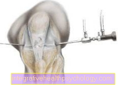
The Arthroscope consists of a pipe (Trocar sleeve) and the optics. Through an approximately 5 mm long skin incision below the Kneecap the trocar sleeve is advanced into the joint with a tip (trocar). The trocar is then pulled back out of the joint through the sleeve. The optic is then inserted into the joint through the sleeve remaining in the joint. In addition, two tubes are connected to the arthroscope. Fluid is introduced into the joint through a hose, the other is used to suck out the fluid.
A second skin incision with a length of approx. 5 mm is necessary for the surgical procedure, through which the small surgical instruments can be inserted into the joint.
Occasionally, a separate supply line for fluid, a so-called irrigation cannula, is introduced into the joint through a third small skin incision about 5 mm long above the kneecap.
The optics
Arthroscopic optics consist of a lens system, a light source and a Fiber optic cable. Video cameras in the smallest version and weighing less than 30 g make it possible to record the inside of the joint and to reproduce it enlarged on a screen (monitor). The surgeon no longer has to look inside the joint through the arthroscope, but can work with a view of the monitor (video arthroscopy). The Video technology is more complex. However, it has the advantage that the greater distance between the surgeon and the knee joint greatly reduces the risk of inflammation of the knee joint by germs. In addition, this technology allows the patient to follow the operation if desired and the findings and the operation to be documented.
The supply of liquid
In the normal state, the interior of the joint between the joint capsule and the bony structures is only a narrow gap. It therefore offers little space for the examination and the surgical procedure. For the Arthroscopy is therefore the joint with fluid (for example with physiological saline solution) or in rare cases with gas filled up. This allows a good view of the individual structures.
Rinse (lavage)
A one-time filling of the joint is not enough to achieve good visibility over the long term. Usually the joint needs to be irrigated continuously during the procedure. It is operated under water, so to speak, like in an aquarium.
With the rinse, for example, remains of disintegrated cells (cell debris) and small pieces of cartilage can be removed. This can already bring about a reduction in pain.
Surgical instruments
On the one hand, arthroscopic surgery for meniscus damage is carried out mechanically and / or motor-driven with the smallest surgical instruments specially developed for arthroscopy, which enable touching, cutting, punching, grasping and sucking.
On the other hand, experienced surgeons can also use laser beams to remove meniscus tissue. A study from 1996 came to the result that - with regard to the functionality of the knee joint after the operation - the complex laser arthroscopy is not superior to the mechanical arthroscopy.
Laser surgery has largely been abandoned by leading experts because of the dangers involved in treating cartilage damage and because of the time it takes.
Preparation for the arthroscopic surgery
The Arthroscopy can in general anesthetic, in Regional anesthesia (Epidural / epidural or Spinal anesthesia) and in rare cases also in Local anesthesia (Local anesthesia).
Many surgeons prefer general anesthesia for the following reasons:
- It causes complete relaxation of the Musculature.
- Applying a tourniquet is possible.
- The treatment can be done with less time pressure.
- Number and location of access to knee can be chosen freely.
The same applies to spinal or epidural anesthesia. In addition, the patient can follow the operation here. Often it is not possible to go home without problems after a few hours. With spinal anesthesia there is also the risk of prolonged severe headache.
The Local anesthesia is associated with a relatively low risk. fear or slight pain, however, can cause the muscles to cramp. As a result, the surgeon cannot open the joint space sufficiently for working with the instruments. The result is an inadequate view of the joint. Associated with this is the risk of damaging the delicate cartilage.
Risks
Since arthroscopy a a surgical intervention is the one with a Anesthesia, or partially one anesthesia, are carried out, there are certain risks. Therefore, before the operation, it should be clarified exactly whether the arthroscopy is actually necessary and whether it makes sense in relation to the symptoms and the disease.
Nevertheless, it can be said that arthroscopy through its minimally invasive implementation one of the operations that is one very low risk to recover. There are various studies that indicate a certain probability of serious complications as part of an arthroscopy. This is included depending on the case 1: 10000 or 1: 25000. That means it a serious complication in 0.01% - 0.004% of cases comes. A serious complication in the context of arthroscopy includes one thrombosis, Pulmonary embolism and severe infections (sepsis).
If problems arise, this is usually due to the arthroscopy Injury to the cartilage, cutaneous nerves and smaller blood vessels. The main risk of thrombosis is pulmonary embolism as a secondary disease. The likelihood of this complication is however very low.
Depending on what form of anesthesia and anesthesia is used, there are different risks. Local anesthesia can sometimes be Damage to the cartilage tissue (Chondrolysis) and general anesthesia always involves risks, such as a allergic reaction or aspiration in which the stomach contents enter the windpipe got connected.
It should also be noted that individual closed seasonsprescribed by the doctor must be strictly observed are. If the Joint, relapses may occur possibly a new operation require. If there are allergies, there may also be intolerance to implants that are used during the operation.
A doctor should be consulted, though fever or signs of inflammation after surgery to the affected kneeas well as redness, warming, pain or swelling.
Contraindications
Contraindications to arthroscopy:
Is there a contraindication to the required for this anesthesia (see preparation), then the arthroscopy cannot be performed. A tendency towards embolism and thrombosis can also be a contraindication. Coagulation disorders can occur after the arthroscopy Bruise in the knee lead and must therefore be clarified before the investigation.
An absolute contraindication to arthroscopy, the treatment of choice, exists when local (local) or general (generalized) infections are present. Also prohibits an increased susceptibility to infection, for example under Cortisone or with immunosuppressive therapy, a joint inspection.
-> Continue to the topic of an arthroscopy
Arthroscopy of the knee for a meniscus tear
As meniscus will the Cartilage in the knee joint designated. This is not a coherent cartilage, but rather an inner and an outer meniscus each per knee joint.
The knee joint is one of the most popular heaviest stressed joints in the human body, bringing an increased likelihood of one Cartilage damage and wear and tear. However, complaints can also be considered Result of an accident or a sports injury occur and are especially in sports like Soccer very often represented. In this context one speaks of a traumatic cartilage damage.
An arthroscopy of the knee is common in the context of a meniscus tear carried out. In the case of acute injuries, as well as complaints that occur due to increased stress, there is often a Cartilage tear which can be remedied by surgery using arthroscopy. The operation includes the Removal of cartilage fragments and the Smoothing of the so-called cartilage base. This is supposed to eliminate pain caused by destroyed or damaged cartilage. The duration of the procedure depends on the underlying damage to the meniscus (Duration of an arthroscopy on the knee)
Read more on this topic at:
- Arthroscopy knee
- Meniscal tear
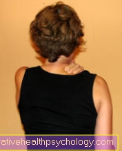
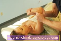
Arthroscopy of the shoulder
Shoulder / rotator cuff
The minimally invasive shoulder surgery Arthroscopy can be useful for various reasons. She's been a popular one for years Alternative to classic surgery on the shoulder as she gentle on the joints is carried out to a significantly faster healing leads and most of all outpatient in a relatively short time can be done.
Most commonly, the shoulder arthroscopy is due to one Joint wear or the so-called Impingement syndrome carried out. If a joint (usually the Shoulder joint, medical: Articulatio acromioclavicularis or AC joint) is affected, the wear and tear on bones and cartilage is repaired by means of surgical equipment during arthroscopy.
The so-called impingement syndrome is caused by the Entrapment of the supraspinatus tendon between two bones (shoulder roof and humerus head). For the stabilization and movement of the shoulder is the Rotator cuff responsible. It consists of four muscles and their tendons in the shoulder area. The Shoulder joint is mainly stabilized by the tendons of these muscles. The tendon of a muscle (Supraspinatus tendon) is however one increased physical stress exposed because it runs in a narrow canal between two bones. In the course of life it can therefore happen that this The tendon wears out and, in the worst case, even tears.
So if there is pain at this point, it is important to have one Check the tear of this tendon. If there is pain in the shoulder area, for example when lying down for a long time on the affected shoulder or lateral lifting of the arm, there may be a torn tendon. To ensure the diagnosis is usually a Ultrasonic carried out, sometimes also a X-ray image be useful for clarification. Usually an arthroscopy of the shoulder follows in order to better assess the damage and - depending on the age and the individual diagnosis of the person affected - to better estimate the further course of action.
In any case, it is important that Customize follow-up treatment of the jointso that the best possible healing is given. Usually one is Load possible again relatively quickly, this can be from a physical therapy or gymnastic exercises.
Read more on this topic at: Arthroscopy of the shoulder
Arthroscopy of the hip
The hip joint belongs to the only recently using arthroscopy treated joints. It was before the introduction of arthroscopy in this area only with the most complex methods possible to carry out small and large repairs on the joint. This led to long rehabilitation periods and one increased number of complications through the operation.
Nowadays the Arthroscopy of the hip for different indications used. Mostly will Cartilage damage and Impingement Syndrome of the Hip, but also light ones Osteoarthritis, free joint bodies or for example a Tear of the headband (labrum) so repaired.
The operation will under general anesthesia carried out. It will, as with most arthroscopies two access routes placed through which surgical instruments can be inserted. Deliver for the positive outcome of the operation mobile X-ray machines precise information about the exact position of the equipment. In order for the surgeon to be able to take a good look at the joint and assess damage properly, it must affected leg put under tension during arthroscopy of the hip become. The surgery can be either lying on your side or on your back respectively.
After an operation it is important that To spare the joint for the time being. Depending on the healing process, full weight bearing can be carried out in the first two weeks after the hip arthroscopy, but it should to unusually high loadsas they arise in sports, for example, be waived. It may also be necessary during this time Forearm crutches to use. A physiotherapy treatment with physiotherapy makes sense because without exercises, there is a risk of restricted mobility.
Arthroscopy of the Elbow
The surgical intervention using arthroscopy of the elbow joint has gained significantly in influence in recent years. As with most arthroscopies, it is performed through two incisions through which surgical instruments can be inserted.
Elbow arthroscopy enables Diagnosis and treatment some painful conditions in the elbow joint. However, other structures in this area can also be examined with the help of arthroscopy. This includes the Endoscopy of the ulnar nerve as well as the Biceps tendon arthroscopy.
Indications there are many for an arthroscopy of the elbow. So be for example Signs of wear and tear on the cartilage, Bursitis, Infections and a Biceps tendon tear treated like that. Even if the pain is unclear, an arthroscopy may be performed to clear it up. The famous Tennis elbow (medical: Epicondylitis), which is based on irritation of the tendon attachments of the muscles of the forearm, can be treated with an arthroscopy.
The procedure lasts after the patient has been prepared, depending on the indication 10 to 60 minutes. The patient lies down during the operation on your stomach, or on your side. For anesthesia, the patient can either use one short general anesthesia, or block anesthesia receive. The joint is not fixed during the entire time, so that the attending physician has the greatest possible overview of the damage.
Overall, the operation is through an arthroscopy on the elbow To be preferred to an open "classic" operation. The surrounding tissue is spared, and the Rehabilitation time shortened. This is why arthroscopy of the elbow is usually the method of choice after classic pharmacotherapy has failed. However, since the structures in the area of the elbow are very close together, there is one steady hand and a trained surgeon essential for the success of the procedure.
Arthroscopy of the ankle
A Arthroscopy of the ankle is a great way of diagnosing and treating some diseases of this region that alternatively only treatable by open surgery would be which with significantly higher risks and rehabilitation times went along. There are different reasons why an ankle arthroscopy makes sense. Through them you can Cartilage damage, free joint bodies, Joint mucosal diseases, Bone spurs and Instabilities of the joint are treated. As with most arthroscopies, two incisions are made that allow the surgical instruments to be inserted. The procedure is under a General or local anesthesia carried out.
After the operation is a Full load in the first two weeks possible after the operation, but things that put stress on the joints such as sport should be avoided. During this time, the Use of forearm crutches be useful. A physiotherapeutic follow-up treatment with physiotherapy exercises prevents permanent restriction of movement.
Read more on this topic at: Arthroscopy of the ankle


.jpg)