Structure of the eye
Synonyms in a broader sense
Medical: Organum visus
Eye structure, eye anatomy, eye
English: eye
introduction
The human eye or the skin of the eye can be roughly divided into 3 layers:
- Outer skin of the eye (dermis and cornea)
- Middle skin of the eye (deer skin, ciliary body, choroid)
- Inner eye skin (retina)
Specialized pigment cells (melanocytes) embedded in the iris (rainbow skin) are responsible for the eye color that is visible from the outside. The amount of pigment alone determines the color of the eyes: brown eyes contain a lot of pigment, while blue and gray eyes contain little.
Belonging to the middle skin of the eye (the so-called tunica vasculosa bulbi, the vascular skin), the iris borders on the back skin of the eye, the retina. In addition, the radiation bodies (lat. corpus ciliare, Ciliary body) and the choroid, which supplies the outer retina with blood (Choroid) to the middle skin of the eye.
Another important function of the radiating body is the formation of the aqueous humor. This structure is also used to attach the lens, which is hung on straps behind the iris. The entirety of the structures belonging to the middle skin of the eye is also known as the uvea.
The Lens
In addition to the cornea, the lens is the second light-refracting, transparent organ in the eye. In contrast to the latter, however, its refractive power is variable, so that a sharp image of near and distant objects on the retina is possible.
Responsible for this is the muscular length of the suspension straps of the lens: If they slack, the lens bends passively due to its inherent elasticity and the refractive power increases: nearby objects can be seen clearly with the eye. If the suspension straps are tightened, the lens becomes flatter again as the refractive power decreases. If the ratio of the lens refractive power does not correspond to the length of the eyeball (i.e. the distance from the retina), a sharp image cannot be produced on the retina.
These eye diseases (Ametropia) are corrected by increasing or decreasing the refractive power of the lens: In the case of farsightedness (hyperopia), the light is bundled behind the retina, corresponding to a too low refractive power of the eye or a too short eyeball. Therefore, this construction, a converging lens that focuses the light (with positive refractive power; this is measured in dioptres) can help here. In myopia, the refractive power of the eye is too great or the eyeball too long and the sharp image is shown in front of the retina. The treatment is therefore carried out with diffusing lenses (with negative refractive power).
More on this: Lens of the eye
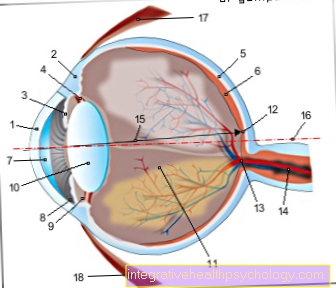
- Cornea - Cornea
- Dermis - Sclera
- Iris - iris
- Radiant Bodies - Corpus ciliary
- Choroid - Choroid
- Retina - retina
- Anterior chamber of the eye -
Camera anterior - Chamber angle -
Angulus irodocomealis - Posterior chamber of the eye -
Camera posterior - Eye lens - Lens
- Vitreous - Corpus vitreum
- Yellow spot - Macula lutea
- Blind spot -
Discus nervi optici - Optic nerve (2nd cranial nerve) -
Optic nerve - Main line of sight - Axis opticus
- Axis of the eyeball - Axis bulbi
- Lateral rectus eye muscle -
Lateral rectus muscle - Inner rectus eye muscle -
Medial rectus muscle
You can find an overview of all Dr-Gumpert images at: medical illustrations
The retina

The structure of the back wall of the eyeball is lined on the inside by the retina. It mainly consists of nerve cells that are responsible for converting light stimuli into electrical signals and transmitting them to the brain. This section of the eye, also known as the fundus of the eye, is accessible to the medical examination by looking through the medically dilated pupil (Fundoscopy).
The most important structures are:
- blind spot and the
- yellow spot (Latin macula lutea).
The blind spot is the place on the retina where the bundled fibers of all nerve cells unite to form the optic nerve (hence the Latin name discus nervi optici). There are no nerve cells there that are necessary for the visual process. Nevertheless, the blind spot is not noticeable as a visual field loss: The missing optical information is controlled by the brain and replaced by the other eye.
On the other hand, the nerve cell density is particularly high at the yellow spot:
This is why it is also known as the “point of sharpest vision. Therefore, z. B. age-related changes have a particularly strong effect on the eyesight (see diseases: age-related macular degeneration). The so-called visual pigment (visual pigment) is important for the visual process. It lies in the processes of nerve cells called photoreceptors and changes its chemical structure when the eye is illuminated, generating electrical signals. For this process, known as transduction (conversion), vitamin A is necessary because it is part of the visual pigment. In the case of vitamin A deficiency, night blindness occurs (Hemeralopia). You can find out more about this disease under night blindness.
The lid, which is one of the auxiliary structures of the eye, is made up of the facial nerve (lat. Facial nerve) controlled (innervated).
Metabolic processes or injuries that lead to damage to the facial nerves are therefore noticeable through reduced or absent eyelid closure. 30 glands contained in the lid produce a fatty film that protects against evaporation of the tear film and thus prevents the eye from drying out. The tear fluid itself is formed by the lacrimal gland located in the lateral, bony eye socket (orbit) (about ½ ml. Per day).
In addition to water, the most important components are proteins that kill bacteria.


.jpg)