Eye socket
anatomy
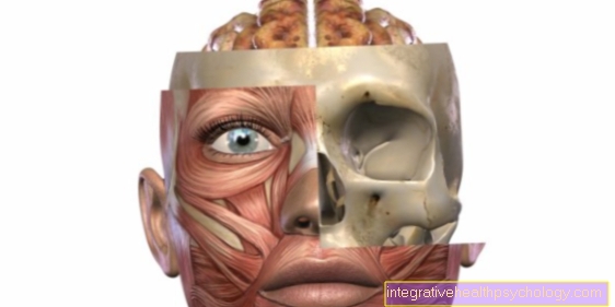
The eye socket, in technical jargon as "Orbit“Is the paired cavity that contains the eyeball and the appendage organs of the visual apparatus.
The bones of the skull are divided into the cranial skull and the facial skull. The facial skull comprises many small bones that form the fine structures in the face and give it its shape.
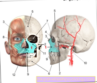
The eye socket is a pit about five centimeters deep, which is formed by seven different bones. The outer edges of the eye socket are easy to feel. The palpable edges are formed in the middle by the upper jawbone, outside by the zygomatic bone and above by the frontal bone.
What is the orbital floor?
The eye socket (Orbit) is a bony structure in which the eye is embedded for protection. It is pyramidal and consists of the orbital roof, the orbital floor and two lateral boundaries.
The floor of the orbit consists of three bones. These include the maxillary bone (Maxilla), the palatine bone (Palatine bone) and the cheekbone (Os zygomaticum). The jaw cavity borders on the floor of the eye socket (Maxillary sinus).
There is also the infraorbital canal in the floor of the orbit. This is a small passage in the bone through which the artery, vena and infraorbital nerve run. These are responsible for the blood supply and sensitivity in the area between the lid and upper lip.
What bones is the eye socket made of?
The eye socket (Orbit) is a bony cavity that surrounds our eye. It is made up of seven bones in total.
The floor of the orbital socket includes:
- Maxillary bone (Maxilla)
- Palatine bone (Palatine bone)
- Cheekbone (Os zygomaticum)
Limits the eye socket from above and is therefore the roof of the eye socket:
- Frontal bone (Frontal bone)
At the side the eye socket is supplemented by:
- Tear bone (Lacrimal bone)
- Ethmoid (Ethmoid bone)
- Sphenoid bone (Sphenoid bone) added.
This bony protective structure for the eye contains numerous through-holes for various vessels and nerves. In addition, the eye socket is filled with fat and connective tissue, into which the eye and other structures (e.g. Lacrimal gland, eye muscles) are embedded.
Function of the eye socket
Their main function is to protect the sensitive human visual apparatus from external violence. The protruding bone edges are therefore the most common location for injuries. The outer opening of the eye socket is filled by the eyeball and the appendage organs. This is covered by the skin of the face and the eyelids, so that only part of the white skin of the eye, the iris and the pupil can be seen from the outside.
In the interior of the skull, the bony border of the eye socket converges in a conical shape. Inside, there are only small holes and channels as access points, in which, among other things, the optic nerve runs.
Normally, around six eye muscles run from the eyeball to the rear edge of the bony eye socket, which hold the eye in its position and enable the eyeball to move. The lacrimal gland is located above the eye and shifted slightly outwards.
Numerous nerves and blood vessels run within the orbit. They supply the structures within the eye socket, for example the eyeball and lacrimal gland.
Some of them also move into the central nose and supply it with blood and sensitive nerves. Individual branches also pull from the eye socket to the front of the face and are responsible for the sensitive sensation from the upper lip to above the forehead.
Failures of conduction pathways within the eye socket are expressed in different ways as loss of sensitivity in the face, as visual impairment or, for example, by seeing double images.
Read more about this at: Injury to the visual pathway
Diseases of the eye socket
Pain in the eye socket
Some of the structures within the eye socket are sensitive to pain and can become ill. Pain in the eye is particularly frequent caused by the eyelids, the lacrimal gland or the conjunctiva.
Since the eye socket offers an opening into the interior of the body, it is also a gateway for pathogens that can cause painful inflammation.
One of the most common causes of blindness worldwide is this glaucoma. Glaucoma, which is also known as glaucoma, leads to high pressure in the eye and can occur as an attack with severe eye pain.
Read more on this topic: Emergence of glaucoma
Less common causes of acute pain in the eye socket can be injuries, for example foreign bodies in the eye, broken bones in the eye socket or chemical burns. A tumor as a pain-causing foreign body can also be the cause.
Read more on this topic at: Pain in the eye socket
Orbital hernia
An orbital hernia is also known as Orbital fracture designated. It occurs largely as a result of blunt violence, for example falling on the face, colliding with solid objects or as a result of conscious violence (blows).
More information is available here: Orbital hernia
The bony structures of the eye socket are supposed to protect the eye from that kind of violence. If the external bones are bruised, the eye socket can break. In most cases, the fracture occurs either in the floor or roof of the eye socket, with the orbital floor fracture being more common.
The secondary consequence is visual impairment. Double vision and restricted eye movements are the most common consequences.
There may also be bruises in the eye socket. The intraocular pressure can also increase. If the sensitive nerves are affected, tingling and impaired perception can occur in the facial area.
With the help of certain eye tests, doctors can determine whether the problem is short-term failure of the eye muscles or actual paralysis. Often an improvement occurs on its own after a few weeks. Surgery for orbital fractures is very controversial as the success of interventions is only moderate.
The severity of the symptoms and the subsequent treatment depend on the severity of the fracture. Often only one wall is affected, but in severe comminuted fractures up to four walls of the eye socket can be broken.
Read about this too: Cheekbone fracture
Swelling in the eye socket
Swelling in the eye socket can have several causes. An easily treatable cause is inflammation, which starts from e.g. the sinus or an inflamed tooth into the eye socket.
This swelling is treated with antibiotic therapy, which should be started as soon as possible.
An endocrine orbitopathy is also possible (EO), which in most cases occurs in connection with Graves' disease. It is an autoimmune disease that causes enlargement of retrobulbar structures (Connective, fat and muscle tissue) leads.
A more serious disease is rhabdomyosarcoma, which often manifests itself in the eye socket. This is a malignant tumor that is treated with an operation and subsequent radiation or chemotherapy.
You can find more interesting information on this topic at: Swelling of the eyes
Inflammation of the eye socket
The most common cause of eye pain is inflammation of the structures of the eye socket. Bacteria or viruses are often responsible for the inflammation, but sometimes also fungi or parasites. The eye fends off a large part of the pathogens every day, but it always offers a potential portal of entry into the body.
In particular, smear infections caused by one's own hand lead to inflammation. External stimuli such as bright sunlight, dust or permanent drafts on the eye can also cause inflammation.
Theoretically all structures of the eye socket can be affected, the lids, the lacrimal glands, the cornea, the outer, middle and inner eye skin, but also the optic nerve or the eye muscles. Especially conjunctivitis, the so-called "Conjunctivitis“Is a typical clinical picture. Symptoms are externally visible redness, sensitivity to touch, feelings of foreign bodies and sometimes purulent secretions with sticking of the eyelids.
Read more about this: Symptoms of conjunctivitis
In very rare cases, the so-called "Trigeminal nerve " come, whereby even light touches of the facial skin can cause stabbing pain.
Read more about this: Trigeminal neuralgia
MRI of the eye socket
Imaging in diseases of the eye socket is of great importance. In particular the MRI (Magnetic resonance imaging) represents the eye socket and the surrounding soft tissue (Connective tissue, muscle tissue and underlying structures such as nerves and vessels) very well.
It is best suited for inflammatory and neoplastic processes in the eye socket, as it enables the highest possible contrast. In addition, the MRI works completely without harmful radiation, which is another advantage for the patient.
One situation where MRI is less suitable is any type of emergency situation. Even if an injury to the eye socket is suspected, e.g. after a high-speed trauma, the CT (Computed Tomography) is preferred because it can generate an image in a much shorter time.
Read more about MRI under: Clothes in the MRI - what should I take off, what should I wear?