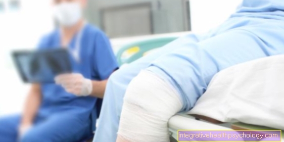
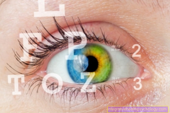
Fundus examination
Synonyms in a broader sense
Control of the fundus, observation of the retina, retinal mirroring, funduscopy, ophthalmoscopy
What is the investigation for?
An examination of the fundus is usually not necessary as long as the patient does not have any symptoms and there have never been any problems with the eye and especially with the fundus in the past.
The examination of the fundus is an informative and important examination, as many diseases can be identified and controlled through it. How often the patient should come for an ocular fundus examination depends entirely on the individual case and must be decided by the attending physician.
Both the retina and the optic nerve head (papilla) can be viewed and important supplying structures, such as the blood vessels, but also the point of sharpest vision (Fovea) can be assessed.
The fundus of the eye should not be confused with the intraocular pressure measurement, which must be paid for by the patient himself and which is nowadays suspected of being carried out too often unnecessarily and without diagnostic benefit.

Basic information about the investigation
In the examination falls on the Retina a relative strong lightwhat glare-sensitive people can find disturbing.
Of the Examination room yourself should darkened be so the Ophthalmologistwho carries out the fundus, is not blinded from the outside during the examination and the examined structures can be better recognized and assessed.
To the pupil to expand and thereby one better insight on the Fundus to achieve will mostly dilating eye drops given.
This gift is in the most cases without any other side effects.
However, it should be ensured beforehand that the patient's anterior chamber is not too shallow, since the intraocular pressure can increase due to the dilated pupil and there is thus the risk that the patient will suffer from it acute attack of glaucoma comes. This increases the Intraocular pressure strong, there is pain and there is a risk of permanent Visual impairment.
Since the Fundus examination however usually taking place in an ophthalmologic clinic or practice, the patient becomes the Working time the drop over supervised and if the typical symptoms occur, appropriate counter-therapy is initiated.
It is important that the patient with the pupils enlarged by the drops do not operate a vehicle may. The patient is only allowed to drive again when the effect has subsided a few hours after the fundus examination.
If the use of the eye drops is not possible, the fundus examination can still be carried out: The ophthalmologist then only has a limited view of the fundus and can judge subtleties and details more poorly due to the restricted illumination. However, this is often sufficient to get an orientation overview of the condition of the fundus.
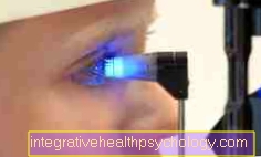
Direct mirroring

The direct reflection of the fundus (direct ophthalmoscopy) generates a upright image. The holds Investigator at a short distance from the eye electrical viewing device. You may have to Corrective lenses can be switched on in the electric ophthalmoscope (Rekoss disc) to a clear image to see. These lenses are also required for spatial measurement.
In direct ophthalmoscopy (Retinaloscopy) becomes a strong magnification causes, however, only one small part of the fundus can be seen.
Since an upright image is created here, even an inexperienced examiner can assess the findings more easily.
Indirect mirroring
Most ophthalmologists use the indirect reflection the fundus preferred. The examiner lights up with a Pupil light the area of the Fundus out. The other hand holds one Magnifying glass in front of the eye of the patient, giving the doctor a mirrored and upside down image sees.
At the doctor's command, the patient looks in different directions so that the doctor can see and assess the different areas of the retina.
There are also special devices, with whom instead of with one eye, with both eyes can be examined and such a three-dimensional image the structures of the fundus can be seen.
With indirect mirroring (indirect ophthalmoscopy) large areas can be viewed and such a Overview about the Retina to be obtained.
Duration of an ocular fundus examination
A fundus examination is part of the ophthalmological examination routine and does not take much time in itself. However, since the pupils of the eyes have to be artificially opened using anticholinergic eye drops before the actual examination, it is necessary to allow a little more time.
The patient is often given the drops by a nurse and asked to sit down for a few minutes until the eye drops take their full effect and the pupils are completely open. Then the real investigation can begin.
Depending on which of the types of examination the doctor decides and what he considers necessary, the fundus examination takes between five to ten minutes. It is of course possible that the ophthalmologist will discover abnormalities that require further examinations afterwards, which can then take longer. When the examination of the fundus is completed, the patient can in principle walk again immediately. However, he should bear in mind that the pupil-expanding effect of the eye drops can last up to five hours and that he is therefore not allowed to actively participate in public traffic during this time.
Which changes can be examined by mirroring the fundus?
Important examples:
- glaucoma (Pressure damage to the optic nerve head)
- Congestive papilla (Swelling of the optic nerve head due to increased intracranial pressure)
- Changes due to diabetes mellitus (Diabetes)
- Changes caused by high blood pressure
- Vascular occlusions (Vein occlusions, Arterial occlusions)
- Retinal holes, tears
- Tumors in the eye
- Retinal detachment
and other abnormalities and diseases.
Fundus examination in children
In the case of healthy children born in the normal way, an examination of the fundus is usually not necessary. However, this is at Premature babies or babies who are born with it Complications came and followed for a while artificially ventilated had to be different. The reason is that the small blood vessels in the retina of the eye are not fully formed until the end of the ninth month of pregnancy and can thus optimally supply the retina with blood and thus with oxygen. If a child is born too early and may need artificial ventilation, this can lead to an excessive formation of blood vessels in the retina, the so-called Retinopathy of premature infants. The more immature the premature baby, the greater the risk.
Retinopathy of premature babies can be diagnosed with the help of a Fundus examination, during which the doctor will check the condition of the retina and assess the development and formation of the blood vessels. If a baby is born too early and consequential damage to the fundus is to be expected, there will be several immediately Control appointments agreed, in which the fundus must be checked. The procedure of the examination is the same as for adults, only with the problem that the little ones cannot cooperate. Therefore, the doctor has to open the eyelids himself and keep them open during the entire examination in order to be able to insert the eye drops and look at the fundus with the magnifying glass. There are also special eyelid locks that push the eyelids apart like a kind of forceps and hold them in this position. Since the baby cannot deliberately look in different directions, but this would be necessary so that the doctor can also see the entire retina, especially the outer areas, he uses a kind of small hook with which he can easily insert the child's eye into the corresponding area Direction pushes. Everything works, thanks topical anesthetic eye drops, is painless for the child, but is still uncomfortable and especially unpleasant for the parents to watch. The check-ups will continue until the doctor is of the opinion that the blood vessels have stopped growing and there is no longer any risk of developing retinopathy of premature babies.
Fundus examination in diabetes
Of the Diabetes mellitus is a disease that primarily affects the pancreas and thus the body's sugar metabolism. However, this disorder affects the entire body and all organ systems, including the eyes.
The main sequelae that diabetes causes in the eye are Developing a cataract, also cataract called that Diabetic retinopathy and the secondary or neovascularization glaucoma.
Diabetic retinopathy, in turn, can be divided into two sub-forms, on the one hand proliferative and on the other hand the non-proliferative form. The blood sugar, which has often been poorly controlled for years, and the too high blood pressure of those affected cause minor damage to the blood vessels in the back of the eye. These supply the retina, which can suffer damage and die without sufficient oxygen. Due to the small lesions that have arisen in the vessel wall, small bulges, so-called bulges, form there Microaneurysmsthat can burst and so for Bleeding in the retina to care. This stage of retinopathy is called non-proliferative. In a more advanced stage it then comes to Formation of new small blood vessels in the retina, as this is dependent on a sufficient supply of oxygen, but the blood can no longer be transported everywhere through the defective vessels. However, these fresh vessels are very weak and vulnerable, which further increases the risk of bleeding into the retina. If the blood even gets into the vitreous humor of the eye itself, vision is severely impaired. Despite the severity of the disease, those affected notice it in the early days no symptoms for a long time. Sometimes eyesight is slightly restricted and vision becomes a little blurry. If the first symptoms then appear, it is usually too late to be able to effectively combat the progression and the consequential damage must first and foremost be treated. Hence it is extremely important for diabetics to be informed by the attending physician regular fundus exams also to be conscientious, as precaution is the best way to avoid damage to the eye.
The cataract is a gradual one Clouding of the lenswhich obstructs the light entering the eye and restricts vision. A cataract in old age, i.e. over 65 years of age, is quite normal. But it often develops a lot in diabetics faster and earlier. The first symptoms can be cloudy and blotchy vision, increasingly poor night vision, light sensitivity and halos around bright light sources. The cloudiness does not cause pain, which is why the creeping process is hardly or only very difficult for many of those affected noticed late.
Proceed further
For some of the diseases, further examinations will then be required, such as one Vascular representation (Fluorescence angiography) or one Investigation with Ultrasonic.
Driving after an ocular fundus examination
Following a fundus examination, the patient should do not drive a car for a period of time and not actively participate in public road traffic in any other way. Because the pupils are dilated with drugs, they are no longer able to adapt to the incident light of the environment and to seeing near and far. The Vision becomes blurred and the eye is more sensitive to light than normal. As a result, you can see much worse than normal and therefore often cannot correctly assess distances and speeds. As a rule, however, the effect of the pupil-dilating eye drops should be gone after about three to five hours and the patient can again take part in traffic without restrictions. The widening of the pupil for the examination remains natural without further consequences for the eye or sight.