Eye muscles
Precise mobility of the eye is necessary for seeing. This is ensured on the one hand by the outer eye muscles and the inner eye muscles are responsible for "focusing".
A third muscle group on the eye, which is partially assigned to the so-called mimic muscles, is responsible for the movement of the eyelid.

Eye muscles anatomy
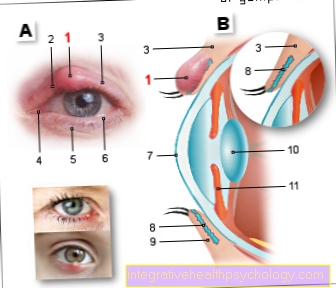
The human eye is able to move and rotate in many different directions. The movements of the eyeball are made possible by a complex interplay of different muscles.
These muscles are called the outer eye muscles because they attach to the outside of the eyeball. The outer muscles of the eye can be controlled consciously and voluntarily. A distinction is made between the outer eye muscles and the inner eye muscles, which are located inside the eyeball and are used for accommodation (change in the refractive power of the eye depending on the distance of the object being viewed) and pupil motor functions (size of the pupil depending on the light conditions in the environment). These inner eye muscles cannot be consciously controlled.
An optimal interaction of all external and internal eye muscles is of great importance for sharp vision. Damage to just one muscle can lead to double vision, blurred vision and squinting.
Course and function of the eye muscles
The external eye muscles
The outer eye muscles, which are used for the conscious and voluntary movement of the eyeball in different directions, consist of six eye muscles, the four straight (Latin: rectus) eye muscles Superior rectus muscle, Inferior rectus muscle, Medial rectus muscle and Lateral rectus muscle, as well as the two oblique (Latin: obliquus) eye muscles Superior oblique muscle and Inferior obliquus muscle.
The four straight muscles of the external eye muscles have their common origin in a ring-shaped optic plate, the so-called Anulus tendineus communis, which is located deep in the eye socket and has grown there with the bone. From here the straight eye muscles pull forward to the eyeball.
The superior rectus muscle extends straight forward from the common tendinous annulus and attaches to the top of the eyeball. When contracted, the superior rectus muscle moves the eye upward (superior) and inward. At the same time, the superior rectus muscle can cause the eyeball to roll inwards.
The rectus inferior muscle also pulls straight forward from the anulus tendineus communis, but attaches to the bottom of the eyeball and therefore moves the eye downwards (inferiorly) and inwards when tensed. At the same time, the inferior rectus muscle can cause the eyeball to roll outward.
The medial rectus muscle pulls straight forward from the anulus tendineus communis and attaches to the inside of the eye, i.e. the nasal part of the eye (medial) and moves the eye inwards towards the nose when it is tensed.
The rectus lateralis muscle, on the other hand, pulls forward from the anulus tendineus communis to the outside of the eyeball and moves the eye outwards (laterally) when it is tensed.
The superior oblique muscle has its origin in the upper inner (nasal) part of the eye socket and moves forward from there. After a short course through the eye socket in the direction of the eyeball, the superior oblique muscle is deflected on a roll-shaped cartilage, the so-called trochlea, and now runs outwards instead of forwards. Finally, it starts at the top, the outside and the back of the eyeball. Due to this complex course, the superior oblique muscle is able to roll the eye inwards, as well as to move the eye downwards (lowering) and outwards.
The inferior oblique muscle, on the other hand, has its origin in the lower inner (nasal) part of the eye socket. From here it runs beneath the inferior rectus muscle through the eye socket and finally starts at the bottom, outside and back of the eyeball. If the inferior oblique muscle is tightened, this causes the eye to roll outwards and to move the eye upwards (lifting) and outwards.
The inner eye muscles
The inner eye muscles that the Accommodation (Change in the refractive power of the eye depending on the distance of the viewed object) and the Pupillomotor function (Size of the pupil depending on the light conditions in the environment) is formed by three muscles, the ciliaris muscle, the sphincter pupillae muscle and the pupillae dilatator muscle.
The Ciliary muscle arises from a layer that surrounds the eye from the outside, protects it and, among other things, serves to shape the eyeball, the so-called sclera or dermis. The ciliary muscle is connected to the so-called zonular fibers, which in turn are connected to the lens of the eye. If the ciliary muscle is relaxed, the zonular fibers are taut and pull the lens flat. When the ciliary muscle tenses, the zonular fibers relax and the tension on the lens is released, causing the lens to twist. Depending on the tension of the ciliary muscle, the shape of the lens changes.
The change in shape of the lens also changes the refractive power of the lens, which is known as Accommodation designated. Through accommodation, which happens unconsciously and involuntarily, it is possible for us to see clearly objects that are near as well as far away from us.
The Sphincter pupillae muscle and the Dilator pupillae muscle are ring-shaped muscles that surround the pupil in a circle.
The Sphincter pupillae muscle causes a reduction in the size of the pupil, the Dilator pupillae muscle on the other hand, an enlargement of the pupil.
These two muscles are important in regulating the amount of light entering the eye.
If the area around the eye is very bright, for example in strong sunlight, the sphincter pupillae muscle causes the pupil to constrict and thus prevents too much light from entering the eye and blinding you. In contrast to this, the dilatator pupillae muscle causes the pupil to widen in weak light conditions, for example at dusk, so that more light enters the eye and one can see despite the dusk. These two muscles cannot be consciously controlled either.
The muscles of the eyelid
Muscles that are able to move the eyelid are the Levator palpebrae superioris muscle and the Orbicularis oculi muscle.
The Levator palpebrae superioris muscle arises like the straight outer muscles of the eye Anulus tendineus communis (a tendon ring in the eye socket) and pulls forward through the eye socket into the upper eyelid, in which it expands in a fan shape.
The levator palpebrae superioris muscle can open and retract the eyelid and is therefore also known as the eyelid lifter. The orbicularis oculi muscle surrounds the eye in a circular manner and causes the eyelid to close when it is tensed.
Innervation of the eye muscles
In order for eye muscles to tense up and move, they need signals (commands) from nerves in the brain. An important nerve that transmits such signals to the muscles of the eye is the oculomotor nerve. It supplies most of the outer muscles of the eye, the superior rectus muscle, the inferior rectus muscle, the medial rectus muscle, the inferior oblique muscle, and a muscle that is responsible for lifting the eyelid, the levator palpebrae superioris muscle.
Another important nerve for the external eye muscles is the trochlear nerve, which supplies the superior oblique muscle. The lateral rectus muscle, which also belongs to the external eye muscles, is supplied with electrical signals by another nerve, the abducens nerve. The orbicularis oculi muscle receives the signals from the facial nerve, which also supplies many other muscles in the face. Musculus ciliaris, Musculus sphincter pupillae and Musculus dilatator pupillae receive their signals via the so-called autonomous nervous system. This autonomous nervous system consists among other things of the sympathetic and the parasympathetic and cannot be consciously controlled.
Blood supply
In addition to an electrical signal, which they use various annoy received, the eye muscles also need one Supply of blood, in order to work and Perform movements. Branches of the are essential in the blood supply to the eye muscles Ophthalmic artery involved which a branch of Internal carotid artery, the internal carotid artery is.
Diseases of the eye muscles
Inflammation, injuries as a result of accidents, tumors or circulatory disorders can damage the eye muscles and their supplying nerves or blood vessels, which can lead to a loss of function of the affected muscle. Symptoms that can occur when only one muscle is damaged are, for example, double vision, blurred vision or strabismus.
Muscle twitching of the eye
Eye twitching is a common symptom of fatigue, stress or a magnesium deficiency. It usually lasts for a certain time, is only localized in one eye and disappears again by itself. Certain muscles in other parts of the body can also twitch when stressed. However, such twitches are more likely to be noticed in the eye, as the muscles there are pretty much directly under the skin.
If the twitching of the eyes occurs only sporadically, no further investigation is usually necessary. However, if it lasts longer or spreads, a doctor should be consulted to rule out more serious causes. An ametropia, for example, can lead to overexertion of the eye muscles, which in turn can be expressed by twitching. In very rare cases, eye twitching can also be a symptom of a nerve or brain disease or a tumor.
Read more on the subject Twitching eyes.
What is eye muscle paralysis?
Eye muscle paralysis refers to permanent or temporary paralysis of one or more eye muscles that can occur on one or both sides. As a result, the movement of the two eyes is no longer coordinated with each other and visual disturbances occur.
This paralysis can have several causes. All causes are serious illnesses and in some cases can result in permanent damage.
The most common cause of eye muscle paralysis is a stroke, which damages some areas of the brain. The cranial nerves that innervate the eye muscles can also be damaged or inflamed.
Furthermore, external forces or trauma can damage the muscles or nerves.
In addition, some systemic diseases such as inflammation of the thyroid gland, inflammation of the muscles or autoimmune diseases such as myasthenia gravis can be the reason for eye muscle paralysis.
Typical symptoms of eye muscle paralysis are
- sudden double vision,
- Dizziness,
- Headache or also
- the drooping of the upper eyelid.
The visual disturbances can also lead to frequent bumping into objects or fine mechanical problems.
If you notice the symptoms described in yourself or others, we urgently recommend that you consult a doctor.
Therapy consists first of all in treating the underlying disease. However, if there is no improvement, after a while you can try to correct the visual disturbances by taking corrective measures on the eye or using glasses. Until this is achieved, no potentially harmful activities such as driving should be carried out.
Find out more about the Eye muscle paralysis.
What is eye muscle inflammation?
Inflammation of the eye muscles, known as myositis, is a rare disease.
The symptoms of ocular muscle inflammation usually go hand in hand with many other symptoms or precede them. It comes to
- Muscle pain,
- Muscle weakness with double vision,
- Dizziness and headache.
In addition, there are often swallowing difficulties, generalized muscle weakness, gait disorders or breathing problems.
The causes of eye muscle inflammation are diverse and can be caused by bacteria, viruses or parasites. Hereditary forms also occur. Inflammation of the eye muscles can also be toxic and result from medication. Another rare cause is autoimmune diseases.
The diagnosis is relatively difficult to make and requires a variety of specific methods, which is why the diagnosis is often made late.
Therapy should be aimed at suppressing inflammation by inhibiting the immune system. If this succeeds, the symptoms usually improve quickly.
Find out more about the Eye muscle inflammation.
Eye muscle spasm
An eye muscle spasm describes a disease in which one or more eye muscles contract permanently and thus do not allow correct movement of the eyeball.
This manifests itself in double vision, pain and other visual disturbances.
The causes for this are varied and not always directly recognizable. For example, a mass in the eye socket such as a tumor, a vascular sac or bleeding from damaged cranial nerves can lead to a spasm of one or more muscles. Degenerative or autoimmune diseases such as multiple sclerosis can also cause eye muscle spasms.
What is an eye muscle weakness?
Eye muscle weakness is an incomplete paralysis of the muscles, which is accompanied by reduced or weakened strength of the eye muscles.
(Eye) muscle weakness is called paresis in medicine and must be differentiated from complete paralysis. In paresis, the function of the antagonistic muscle predominates, whereby the function of the original muscle is either weakened or completely eliminated.
Overall, the clinical picture of weakness of the eye muscles is rare and actually less common than complete paralysis of the eye muscles.
Double vision, dizziness and headache develop. In addition, gait disorders or impaired fine motor skills often occur. If the muscles of the eye are weak, not all, but rather one or a few muscles are usually affected.
The number can depend on the cause. If you consider an eye muscle weakness caused by a cranial nerve, it depends on which cranial nerve is affected. In the event of inflammation or as a result of trauma, several eye muscles are usually affected by an eye muscle weakness. If a rare autoimmune process is the cause, all eye muscles often show weakness.
The therapy for the weakness of the eye muscles depends first of all on the treatment of the underlying disease. However, if there is no improvement, after a while you can try to correct the visual disturbances by taking corrective measures on the eye or using glasses.
Pain in the muscles of the eyes
Pain in the eye muscles is not common.
The symptoms that occur with the pain depend heavily on the underlying clinical picture and can be very unspecific. For example, as with eye muscle inflammation, this can be double vision, dizziness and head jokes. However, visual disturbances, redness, swelling or other pain also occur. Nevertheless, the pain in the eye muscles is usually permanent.
The cause of the pain in the eye muscles can often not be clearly identified, as there are many possible causes. Eye muscle pain can occur in the event of trauma caused by external force, inflammation of the eye muscles, an insufficient supply of oxygen as part of a sinus vein thrombosis, an abscess or tumors in the eye socket or inflammation of the eye socket.
The therapy of eye muscle pain takes place within the framework of the therapy of the cause and can be very different.
Read more about Pain in the eye.
How can you train the eye muscles?
The training of the eye muscles is aimed primarily at people who spend a lot of time in front of the screen and who overstrain their eyes for close viewing over the long term. Here in particular, switching to far-off vision is often neglected. The ring-shaped ciliary muscle in particular is responsible for the different deformation of the lens depending on the distance from the object being viewed. This process is also known as accommodation. When the ciliary muscle contracts, the lens becomes more spherical and nearby objects can be seen clearly. In order to see in the distance, this muscle has to relax and the lens therefore takes on a rather elongated shape.
In people who spend a lot of time in front of the screen, the ciliary muscle is often continuously contracted for very long periods of time. This in turn can lead to myopia in the long term. Eye muscle training should, among other things, start right here and counteract the development of myopia through targeted exercises. For example, exercise packages are offered in which you consciously switch between seeing near and far in order to alternately stress and relax the ciliary muscle. Exercises are also offered against presbyopia, which are intended to counteract the natural stiffening of the lens.
Especially between the ages of 40 and 50, this process can be delayed by a few months or years if the training is well adapted. A complete renouncement of the use of glasses or corrective measures such as laser surgery does not seem to be possible at the moment through eye training.
Are there eye muscle soreness?
There is no sore eye muscles in healthy people. Due to the daily stress, the muscles of the eye are so well trained that they can withstand normal stress.
With some diseases, however, the eye can be misaligned for a longer period of time, which changes the load and gives the feeling of sore muscles. The cause should be treated.
Some other symptoms can also be misinterpreted and attributed to the eye muscles.
If you had an eye muscle spasm, you may experience pain or a feeling of sore muscles in the eye muscles due to the maximum load. However, this feeling should subside after a few days.
Relaxation exercises for the eye muscles
People who spend a lot of time in front of a screen also tend to have tension in the neck and neck area as well as burning or dryness in their eyes. There are also a number of exercises for this that can specifically help to relax the eye muscles. For example, the warm palms of the hands can be placed on the eyes for 10 to 20 seconds by rubbing beforehand or certain points on the bony edge of the eye socket can be massaged with circular movements. On the one hand this can stimulate the blood circulation, on the other hand it helps to release the tension in the muscles. Looking into the distance can also provide relaxation, for example looking out the window every half hour. With your eyes closed you can also look towards the sun. In doing so, however, the head should be turned gradually so that the irradiation does not only occur on one point. The entire exercise should take no more than half a minute.