Baker's cyst in the knee
Synonyms
Popliteal cyst, knee joint ganglion
introduction

Cysts are growths filled with thin or thick contents. These sack-like structures are closed off by a capsule and are usually created by the retention of liquid, i.e. the fluid that forms in the cyst can no longer drain away. A Baker's cyst in the knee is a protrusion of the posterior joint capsule; it is located in the hollow of the knee. One differentiates between a primary and a secondary Baker's cyst. The primary form is mostly present in younger patients and is often idiopathic (cause unknown). The secondary Baker's cyst can result from injuries to the knee joint that are associated with joint effusion. Older patients in particular are affected by this, as they more often suffer from knee problems due to signs of wear and tear. With a Baker's cyst in the knee, the main pain is when the knee is bent.
root cause
As already mentioned, it can be an idiopathic (unknown cause) Baker's cyst (primary Baker's cyst) or it develops from a disease within the knee joint (internal knee disease) with joint effusion. This joint effusion comes about because the body is on Inflammation reacts in joints with increased fluid production (synovial fluid or synovia). The accumulation of fluid in the joint causes a Pressure increasethat ensures that the joint capsule or the inner skin of the joint protrudes backwards into the hollow of the knee. This protuberance is now called Baker's cyst. It is usually located on the inward side of the hollow of the knee, between the tendon attachment of the flexors of the thigh and the upper part of the calf muscle. The access to the cyst - also called the cyst stalk - is narrowed by being trapped in the muscle so that the fluid can no longer flow out of the sac.
Various pre-existing or underlying diseases can cause a Baker's cyst in the knee. These are mostly meniscus damage or lesions. These can be caused by accidents or trauma. A Baker's cyst can also develop as a result of knee osteoarthritis or arthritis. Older people often suffer from osteoarthritis of the knee, as this is the wear and tear on the knee joint structures that are heavily stressed during physical activity. Arthritis is often through rheumatic joint inflammation caused, but can also have a bacterial cause. Any other mechanical irritation of the knee joint (instability of the ligaments, cartilage wear, excessive strain on the knee) can also be the cause. Overloading the knee with subsequent formation of a Baker's cyst in the knee is often the case in young people when these knee joints are involved in strenuous sports. Sometimes you can too Operations cause a Baker's cyst in the knee.
Symptoms
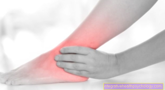
The symptoms of a Baker's cyst in the knee are constant or recurring Pain and a swelling that can be felt to varying degrees in the hollow of the knee in different patients. Furthermore, patients often complain about Feeling of pressure in the hollow of the knee, which is felt as a diffuse pulling / feeling of tension on the back knee when the knee is extended or bent. Depending on the size of the cyst, it happens Restrictions on movement during knee flexion, which increases the feeling of pressure. The pain that is felt increases when the knee is put under strain (for example, when running for a long time or doing sports). The Baker's cyst in the knee can also narrow nerve and blood vessels. Here you can Numbness, Circulatory disorders or Signs of paralysis occur. If the cyst puts pressure on the veins in the legs, the slowing of the blood flow can lead to the formation of a thrombosis (the formation of a blood clot in the vessel).
Appointment with a knee specialist?
I would be happy to advise you!
Who am I?
My name is dr. Nicolas Gumpert. I am a specialist in orthopedics and the founder of .
Various television programs and print media report regularly about my work. On HR television you can see me every 6 weeks live on "Hallo Hessen".
But now enough is indicated ;-)
The knee joint is one of the joints with the greatest stress.
Therefore, the treatment of the knee joint (e.g. meniscus tear, cartilage damage, cruciate ligament damage, runner's knee, etc.) requires a lot of experience.
I treat a wide variety of knee diseases in a conservative way.
The aim of any treatment is treatment without surgery.
Which therapy achieves the best results in the long term can only be determined after looking at all of the information (Examination, X-ray, ultrasound, MRI, etc.) be assessed.
You can find me in:
- Lumedis - your orthopedic surgeon
Kaiserstrasse 14
60311 Frankfurt am Main
Directly to the online appointment arrangement
Unfortunately, it is currently only possible to make an appointment with private health insurers. I hope for your understanding!
Further information about myself can be found at Dr. Nicolas Gumpert
Baker's cyst has burst - what now?
It is rare for the cyst to burst (rupture). If the cyst becomes too large or the pressure created by the fluid increases, the cyst wall can tear - usually when the knee is moved. Often times, patients notice the bursting of the cyst through an increase in pain intensity. If the Baker's cyst ruptures in the knee, it can lead to inflammation in the knee and in the surrounding areas with symptoms of inflammation such as pain, reddening, swelling and overheating. The spread of the inflammation occurs because synovial fluid from the cyst passes into the surrounding tissue. The fluid can spread from the knee to the lower leg and down to the foot. In these tissues there is also an increase in pressure and inflammation, which can constrict and damage surrounding structures such as nerves and blood vessels. This can have serious consequences and should be addressed as soon as possible. It is also important to differentiate the burst cyst from a leg vein thrombosis. This is done by the doctor using duplex sonography (a special ultrasound examination). The blood flow in the veins is shown and a thrombosis is excluded.
What to do?
In most cases, the cyst will resolve on its own. How long this will take and whether it will even happen cannot be said. In the event of pain and restricted mobility, you should consult a doctor. This examines the knee using Ultrasonic and other imaging procedures (MRI of the knee), diagnoses the cause and suggests an appropriate therapeutic measure. In any case, if there is swelling in the back of the knee that is not sure whether it is a cyst, a Exclusion of thrombosis respectively.
therapy
Since the secondary Baker's cyst is always preceded by an inflammatory process in the interior of the joint, the previous illness or the cause of the inflammation should be treated, as otherwise new cysts can form. The cyst usually regresses spontaneously during treatment of the knee joint. The Baker's cyst in the knee can be conservative (without surgery) or with surgery be treated. The operation is usually preceded by a conservative attempt at therapy.
As mentioned earlier, attempts are made to treat the cause in order to cause the cyst to recede. Nonsteroidal drugs are used Anti-inflammatory drugs treated. These include ibuprofen or diclofenac, which have anti-inflammatory and analgesic effects. The attending doctor must decide whether cortisone therapy, which temporarily stops the inflammation process but has some side effects, is necessary. In this case, the cortisone would be injected directly into the knee joint to act against the inflammation on site.
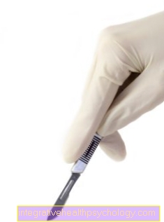
There is also the option of puncturing the Baker's cyst in the knee. However, this procedure is less useful because the likelihood that the cyst will return is very high. If the cyst still does not regress after approx. 6 months, if the swelling is jammed in vascular or nerve tracts, or if the movement is too restricted, the entire cyst can also be surgically removed. This is done through open surgery, in which the cyst is separated from the joint capsule and removed. Furthermore, one should treat the knee damage, if it is present. In the case of meniscus damage, e.g. do an arthroscopy (joint mirroring). The cyst regresses by itself in approx. 60% of cases.
diagnosis
Often times the diagnosis is the Baker's cyst An incidental finding in the knee by the orthopedic surgeon when the knee is examined for other reasons, or by the internist who examines the swelling in the hollow of the knee for thrombosis. After a anamneseA physical exam follows, which reveals medical history and any previous knee problems. The Baker's cyst is usually in the Hollow of the knee felt as a resilient, rounded structure.
Since the Baker's cyst in the knee is usually preceded by a disease of the knee joint, the knee joint should be examined for damage. This can be done by an arthrography (X-ray after previous administration of contrast medium into the joint).
You can also use other imaging techniques such as MRI from the knee or CT the joint and the surrounding soft tissues are shown. The knee damage and also the size of the Baker cyst in the knee and its communication with the knee joint or vessels and nerves can then be assessed. In addition, a Ultrasound examination Thrombosis and Tumors be excluded.
The cause of the Baker's cyst can only be diagnosed with certainty by looking at all possible diagnoses. In many cases it is MRI of the knee joint the most valuable examination method, since one sees Baker's cyst and the causative damage in the knee (e.g. meniscus tear, cartilage damage, cruciate ligament tear) parallel.
forecast
When the Baker's cyst is small, most people have no problem. It often disappears on its own over time or after treatment of the underlying disease. If the cyst becomes larger or if pain, restricted mobility or complications arise, treatment must be given. If you have a chronic disease (rheumatoid arthritis, knee joint disease) that cannot be eliminated, only the symptoms can be treated. If the cyst is surgically removed without treating the cause, it is likely that the cyst will return.
Appointment with a knee specialist?
I would be happy to advise you!
Who am I?
My name is dr. Nicolas Gumpert. I am a specialist in orthopedics and the founder of .
Various television programs and print media report regularly about my work. On HR television you can see me every 6 weeks live on "Hallo Hessen".
But now enough is indicated ;-)
The knee joint is one of the joints with the greatest stress.
Therefore, the treatment of the knee joint (e.g. meniscus tear, cartilage damage, cruciate ligament damage, runner's knee, etc.) requires a lot of experience.
I treat a wide variety of knee diseases in a conservative way.
The aim of any treatment is treatment without surgery.
Which therapy achieves the best results in the long term can only be determined after looking at all of the information (Examination, X-ray, ultrasound, MRI, etc.) be assessed.
You can find me in:
- Lumedis - your orthopedic surgeon
Kaiserstrasse 14
60311 Frankfurt am Main
Directly to the online appointment arrangement
Unfortunately, it is currently only possible to make an appointment with private health insurers. I hope for your understanding!
Further information about myself can be found at Dr. Nicolas Gumpert
Baker's cyst in children
One occurs in children Baker's cyst often in the knee spontaneous and without a known cause. The Baker's cyst in children often remains symptom-free and regresses spontaneously. In some cases, the children sense Feeling of tension and Restrictions on movement - here often after exercise. The Baker's cyst is easy to find with the knee extended or after prolonged physical activity Hollow of the knee Keys. Of course you can also here rheumatic diseases be the cause, but this is rarely the case. A Baker's cyst is also common in children Knock knees on, then on both sides. In any case, the knee and the surrounding soft tissues should also be examined here in order to rule out possible knee joint diseases.
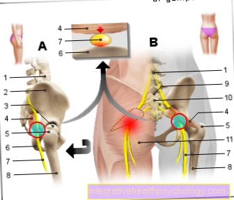
Illustration Baker cyst
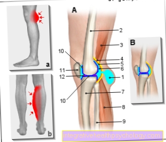
- Baker's cyst
(Popliteal cyst) - Femur - Femur
- Semi-membranous muscle -
Semimembranosus muscle - Joint capsule, fiber layer
(yellow) - Capsula articularis,
Membrana fibrosa - Joint capsule, soft layer
(orange) - capsula articularis,
Synovial membrane - Joint cavity
(filled with synovial fluid) -
Articular cavity, synovia - Shin - Tibia
- Internal calf muscle -
M. gastrocnemius, caput mediale - Fibula - Fibula
- Articular cartilage (dark blue) - Cartilago articularis
- Kneecap - patella
- Inner meniscus - Meniscus medialis
A - Knee joint effusion with Baker's cyst B - Healthy knee joint
a - swelling in the hollow of the knee b - swelling in the calf muscles
You can find an overview of all Dr-Gumpert images at: medical illustrations
prophylaxis
Prophylaxis is hardly possible with a Baker's cyst in the knee, as this is usually a concomitant symptom of a previous inflammatory disease. If the cyst was caused by an inflammation caused by an accident or excessive physical activity and sport, the knee can be spared to reduce the swelling and inflammation. In rheumatic diseases, there are repeated attacks of inflammation, which promote the formation of a cyst again. Therefore, a suitable therapy plan must be worked out with the treating doctor in order to prevent the causal diseases.