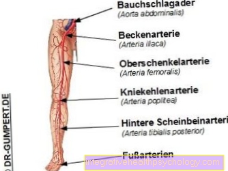
Leg artery
Synonyms
Femoral artery, femoral artery, femoral artery
Also read:
- Types of arteries
- Cardiovascular system
- aorta
- vein
definition
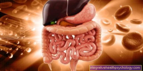
The leg artery (Femoral artery) is the main vessel that supplies the lower extremities with oxygenated blood. In healthy people, it has a diameter of about 1 cm (deviations or differences between the sexes can occur) and gives off numerous branches in its course.
Course of the leg artery
The leg artery (Femoral artery) is symmetrical (i.e. once on both sides). She is the continuation of the External iliac artery (external pelvic artery). The transition from the outer pelvic artery to the femoral artery is located approximately at the level of the inguinal ligament under which the artery runs.
In the course of the leg artery below the inguinal ligament, it is accompanied by the vein of the same name (conduction of oxygen-poor blood from the leg back towards the heart for renewed oxygen loading), i.e. the femoral vein. In the area of the inguinal ligament, there are two important anatomical penetration points for structures that run from the pelvis into the leg. They are Lacuna musculorum (Muscle gate) and Lacuna vasorum (Vascular portal).
To the front (ventral) they are attached to the inguinal ligament (Inguinal ligament) limited, backwards (dorsal) by the pubic bone (Pubis) or iliac bone (Os ilium).
The inside (medial) located vascular port is inwardly through the Lacunar ligament (Gate tape) limited. Through the outside (lateral) Arcus iliopectineus (arched band) it is separated from the muscle port. The arteria and vena femoralis pass through this vascular portal, while the associated nerve (Femoral nerve), accompanied by other structures through which the outer muscle port runs.
The leg artery then runs along the front thigh between the adductor longus muscle and the vastus medialis muscle in the so-called adductor canal. Initially, the leg artery is located relatively centrally on the thigh; as it continues, it pulls more towards the inner front thigh. Through a gap in the adductor magnus muscle (Hiatus adductiorius) the leg artery reaches the hollow of the knee and from there continues as the popliteal artery (Popliteal artery).
Vascular branches
In its course, the artery gives off several vascular branches to supply the surrounding structures:
- The superficial epigastric artery (superficial upper abdominal artery) supplies part of the lower abdominal wall.
- The arteria circumflexa ilium superficialis (superficial artery surrounding the iliac bone) to supply parts of the groin region.
- The external pudendal artery (external pubic artery) for supplying parts of the inguinal skin and the genital area.
- The genicular descending artery (descending knee artery) to the knee joint and
- the arteria profunda femoris (deep femoral artery) to supply the back of the thigh and the femoral head.
Pain
At chronic narrowing of the leg artery due to arteriosclerosis can it to Pain in the calf come. This is due to the fact that there is insufficient blood supply below the constriction. The resulting Circulatory disorder leads to typical symptoms that can be used to trace the narrowing of the leg artery Fontaine-Ratschow divided into 4 stages.
- Stage 1: The vessels are already partially narrowed or even closed. However, the person affected is symptom-free, so that the condition of the vessels is discovered more by chance.
- Stage 2: Kick it Pain when walking on. To relieve the pain, the person concerned stops more often (Intermittent claudication). At this stage, most people see a doctor. One further differentiates:
- Stage 2a: A pain-free walking distance of over 200 m is possible.
- Stage 2b: A pain-free walking distance of less than 200 m is possible.
- Stage 3: Kick it Pain at rest on. Often times, this pain also occurs at night when the legs are horizontal. Letting the legs hang down often relieves the pain symptoms for a short time.
- Stage 4: At this stage it is already closed due to insufficient blood flow Damage to the tissue come (ulcer, gangrene, necrosis). Cell death occurs and toes or other areas of the foot and legs can die. In order to avoid life-threatening situations, appropriate body parts may have to be surgically amputated as an emergency.
Apart from a chronic narrowing of the leg artery due to arteriosclerosis, it can also be caused by a blood clot washed in acute occlusion of the leg artery come, which is represented by severe pain in the leg. The leg subsequently appears pale and cool due to the lack of blood supply and the leg pulses cannot be felt. This represents one emergency and must be treated immediately, otherwise severe damage to the leg can occur.
Narrowing and closure of the leg artery
Constrictions or occlusions in the area of the leg artery can arise suddenly (acute) or over a longer period of time (chronic).
Behind the popularly known "Intermittent claudication"Or the" smoker's leg "hides a chronic narrowing or occlusion of the leg artery. This vascular disease belongs to the complex of peripheral arterial disease.
The cause is mostly a "vascular calcification" (arteriosclerosis), which can lead to a narrowing and eventually also to the occlusion of the leg artery. As a result, the blood vessels behind the narrowed point are not well supplied with blood, the flow rate of the blood is reduced and the arterial blood pressure decreases. Then that Tissue poorly supplied and there are complaints like Pain, feeling cold, paresthesia on the legs (Paresthesia), Skin and nail changes and Skin color changes of the legs.
The body can make use of a number of compensation mechanisms to compensate for the insufficient supply. In addition to improving oxygen uptake and energy generation in the under-supplied leg section, bypass circuits, so-called Collateral circuits form. As a result, the tissue can continue to be supplied with nutrients and oxygen. This Compensations mean that there are only significant symptoms when the closure is around 75%.
In order to determine a chronic occlusive disease of the leg artery, the patient's medical history, including the Risk factors for atherosclerosis recorded. Then the legs are carefully examined. This is followed by palpating the pulses and listening to the leg artery. Also is the Ratschow storage sample carried out in which the patient lies on his back and rides a bicycle with his legs in the air for about 2 minutes. Furthermore, a measurement of the blood flow velocity (Doppler ultrasound examination) and a representation of the vessels with the aid of a contrast agent (Angiography) Provide information about the condition of the leg artery.
In the case of a slight narrowing, in order to prevent further vascular diseases, a Gait training and a Change of lifestyle recommended. If necessary, the therapy can be supplemented with additional medication. Minimally invasive, catheter-based interventions are often performed in the case of medium-severe constrictions. This includes, among other things, the expansion with the help of a small balloon catheter or the removal of arteriosclerotic deposits with a catheter. In the case of severe constrictions and occlusions, an operation on the leg artery is recommended.
From chronic vascular occlusion is the acute occurring Closure of the leg artery to delimit. This emergency caused by blood clots washed in (Embolus) laying the vessel. The place of origin of such an embolus can be the heart. Cardiac arrhythmias such as atrial fibrillation and diseases of the heart valves or valve replacements can cause the formation of a blood clot (thrombus) be. Furthermore, such thrombi can also arise in aneurysms and be washed away.
Characteristic for an acute occlusion of the leg artery are Pain, pallor of the leg that Lack of leg pulses below the closure, paralysis the leg muscles, Sensory disturbances of the leg and finally one State of shock.
After the emergency doctor has established the acute occlusion of the leg artery, an attempt must be made to restore blood flow to the leg as soon as possible. For this you give a high dose of heparin, an anticoagulant, and uses a surgical interventionby removing the occluding blood clot. In addition, one gives strong pain relievers.
Aneurysm of the leg artery
An aneurysm is one abnormal vascular sac of an arterywhich leads to an excessive increase in the diameter of the vessel.
An aneurysm can be congenital or acquired. Of the most important risk factor for the development of an aneurysm arteriosclerosis. This in turn is largely due to obesity, high blood pressure, increased blood lipid levels, smoking and Diabetes mellitus. A bacterial infection can also cause an aneurysm.
If an aneurysm is larger than 3 cm, blood clots (Thrombi) form. This leads to insufficient blood flow in the subsequent tissue sections, which can lead to tingling, numbness and coldness. A thrombus can also be carried away by the bloodstream and move to another location to block a narrowed area (acute arterial occlusion).
If there is no discomfort, an aneurysm is often only found by chance. Imaging procedures such as computed tomography (CT) and magnetic resonance imaging (MRT) are used in the event of complaints.
That is important Treatment of the causes of the aneurysm, e.g. Lifestyle changes are indispensable in the treatment of high blood pressure, if necessary with supportive medication. In other cases, surgery on the aneurysm is recommended. A distinction is made between the insertion of a stent, a so-called Stent, from the formation of a bypass circuit, a so-called bypass.
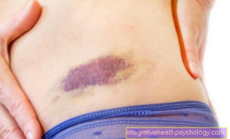
As a complication of a cardiac catheter examination an aneurysm of the leg artery can occur. The reason for this is that for a cardiac catheter examination the leg artery is used as an entry port to the vascular system and is "pricked" (punctured) for this purpose. The resulting damage to the vessel wall can cause it to sag after the examination and form an aneurysm.
Cardiac catheter
To examine the heart with a catheter (left heart catheter examination), the leg artery is mainly used as an access. The catheter, a thin plastic tube, is advanced from the leg artery to the left heart. Contrast media is then injected to show the vessels on an X-ray. Constrictions and occlusions, especially in the coronary arteries, can be detected during a cardiac catheter examination. After assessing the condition of the vessels, a decision can be made about further therapeutic measures.
In some cases, a change in lifestyle, such as diet or exercise adjustments, and drug treatment are sufficient. In other cases it may be necessary to widen the vessels with a small balloon and stabilize the vessels with a stent. These interventions can be carried out directly as part of the cardiac catheter examination.
If the occlusions or constrictions of the blood vessels supplying the heart (coronary arteries) cannot be removed with these methods, an operation may have to be performed (bypass) of the diseased vessels are created.
Read more on this topic: Cardiac catheters

.jpg)


.jpg)