Diagnosing a vertebral fracture
Diagnosis of a vertebral fracture

As always, so also with one Vertebral fracture The physical examination is at the beginning of every diagnosis.
Over one Vertebral fracture can almost without exception trigger pressure and tapping pains. A check of the mobility of the spine should initially not be carried out in order not to provoke fragment displacement in the case of unstable fractures.
There must always be an orientation neurological examination (Sensitivity, voluntary motor skills) in order to provide information on possible injuries to the Backmarks get early.
The conventional one X-ray follows the physical exam. It is advisable to give the indication for the spine exposures generously and not only to x-ray the most painful vertebral body section. In the event of considerable violence, they become one Vertebral fracture (fall injuries, traffic accidents, etc.) is recommended the complete Spine to be examined radiologically. The fear facing the harmful x-rays is almost always exaggerated and unfounded. It can have far more serious consequences if a vertebral fracture is overlooked.
Classic x-rays of the spine are always made in two planes when diagnosing a vertebral fracture, when viewed from the front (a.p.-image) and from the side.
So should be made:
- Cervical spine in 2 planes
- Thoracic spine in 2 planes
- Lumbar spine in 2 planes
Osteoporotic vertebral fracture

Cover and base plate collapse (sintering fracture) in osteoporosis with the formation of short-range kyphosis (rounded back).
If several vertebral bodies break in this way, the so-called "widow's hump" is created, which is characterized by a pronounced rounded back.
Appointment with a back specialist?

I would be happy to advise you!
Who am I?
My name is dr. Nicolas Gumpert. I am a specialist in orthopedics and the founder of .
Various television programs and print media report regularly about my work. On HR television you can see me every 6 weeks live on "Hallo Hessen".
But now enough is indicated ;-)
The spine is difficult to treat. On the one hand it is exposed to high mechanical loads, on the other hand it has great mobility.
The treatment of the spine (e.g. herniated disc, facet syndrome, foramen stenosis, etc.) therefore requires a lot of experience.
I focus on a wide variety of diseases of the spine.
The aim of any treatment is treatment without surgery.
Which therapy achieves the best results in the long term can only be determined after looking at all of the information (Examination, X-ray, ultrasound, MRI, etc.) be assessed.
You can find me in:
- Lumedis - your orthopedic surgeon
Kaiserstrasse 14
60311 Frankfurt am Main
Directly to the online appointment arrangement
Unfortunately, it is currently only possible to make an appointment with private health insurers. I hope for your understanding!
Further information about myself can be found at Dr. Nicolas Gumpert

Most vertebral body fractures / vertebral body fractures can be reliably diagnosed on the X-ray images and an initial assessment of a stable or unstable fracture can be made. If an unstable fracture can be safely ruled out, further diagnostics are not necessary. The typical picture of a stable fracture is the wedge vertebra with a collapsed vertebral body leading edge and intact vertebral body trailing edge. Unstable fractures are not always immediately recognizable in the X-ray image (example of an unstable fracture: see adjacent figure).
If an unstable fracture is suspected, further clarification is required. The extent of the fracture of a vertebral body and thus an assessment of the stability is better than with any other imaging method by means of a computer tomography (CT) (see figure).
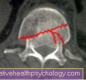
CT of the thoracic spine / lumbar spine
When diagnosing vertebral fracture using Computed Tomography X-ray slices of the vertebral body are made. The course of the fracture can be determined exactly and thus the key question of the involvement of the posterior edge of the vertebral body can be assessed. If the posterior edge is involved in the fracture, the fracture is considered to be unstable and therefore critical to break into the spinal canal.
Here is the particular danger for the spinal cord with a paraplegia.
MRI of the thoracic / lumbar spine
A M.agnetrresonancetomography (MRI) brings with the osseous fracture assessment no further information gain. The bone cannot be assessed so well in an MRI of the transition from the thoracic spine to the lumbar spine. The soft tissues, on the other hand, can be seen much better than on CT.
A MRI of the thoracic spine / MRI of the lumbar spine remains specific questions e.g. after intervertebral disc (Spinal cord) or Soft tissue injuries (Muscles, ligaments etc.) reserved.
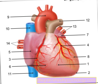
Figure vertebral fracture

Vertebral fracture (vertebral fracture)
- Transverse process -
Transverse process - Spinous process -
Spinous process - Upper articular process -
Superior articular process - Lower articular process -
Inferior articular process - Spinal nerve -
Spinal nerve - Spinal cord -
Medula spinalis - Gelatinous core - Nucleus pulposus
- Vertebral arch - Arcus vertebrae
- Fiber ring - Annulus fibrosus
- Vertebral bodies - Corpus vertebrae
- First thoracic vertebra -
Vertebra thoracica I - Twelfth thoracic vertebra -
Vertebra thoracica XII - First lumbar vertebra -
Vertebra lumbalis I - Fifth lumbar vertebra -
Vertebra lumbalis V
a - cervical spine (cervical spine)
b - thoracic spine (BWS)
c - lumbar spine (lumbar spine)
A - vertebral fracture (spinous process,
Vertebral bodies) from above
B - vertebral fracture (spinous process,
Vertebral body) from the right
C - Most common area of the
Vertebral fracture
You can find an overview of all Dr-Gumpert images at: medical illustrations


.jpg)