Diagnosis of thyroid cancer
diagnosis
At the beginning of the doctor's contact, the patient is asked about his medical history (= anamnesis).
It is of interest here whether the thyroid has changed in size, whether there are swallowing difficulties or a lumpy feeling in the throat.
It is important to find out whether there is a family history of thyroid disorders such as an organ enlargement (= goiter), a Hyperthyroidism, Hypothyroidism or autoimmune diseases that are genetically inherited and lead to the development of thyroid cancer (e.g. MEN = multiple endocrine neoplasms).
The doctor also asks about the patient's medication and whether he has been giving contrast media in the past few months. Contrast media containing iodine can cause a Hyperthyroidism (Hyperthyroidism) and can be problematic for further diagnostic procedures (see scintigraphic examination).
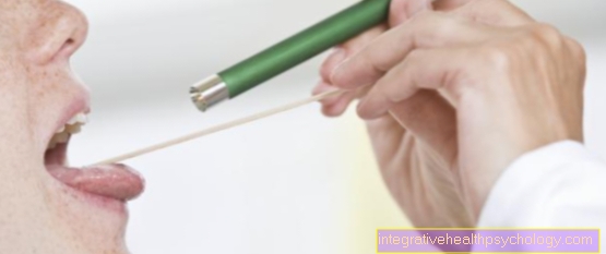
The doctor examines the patient's thyroid while sitting:
The neck is viewed and checked for an enlarged thyroid gland. The thyroid is scanned in a subsequent step.
A detailed physical examination of the patient follows.
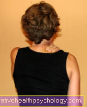
Patients with abnormal thyroid palpation, i.e. those patients in whom one or more Lump in the thyroid gland are noticeable during the palpation examination of the neck, are asked about family diseases:
Note: abnormal palpable finding
Every noticeable palpable finding of the thyroid is clarified in more detail by an ultrasound examination. If there are one or more nodules in the thyroid tissue that are blurred and are hypoechoic in the ultrasound image, a malignant process is suspected.
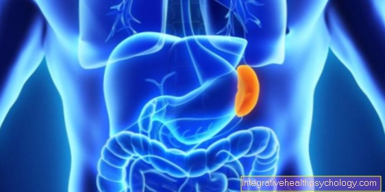
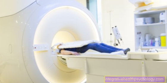
The next diagnostic step is a scintigraphic examination of the thyroid tissue in order to be able to assess the activity of the organ and especially the functional state of the nodular areas.
Functionally active, hormone-forming thyroid cells store iodine.
For the Scintigraphy if you take advantage of this property:
The patient receives iodine through a venous access that is loaded with the radioactive marker 99mTechnecium-Pertechnate. The iodine accumulates together with the technecium in the thyroid tissue, which enables the examiner to make a quantitative statement on thyroid function. A so-called cold lump, which is typically found in a cyst or thyroid cancer, does not store iodine and therefore does not indicate any radioactivity.
Is the cold knots If the ultrasound examination is not echo-free, a malignant thyroid tumor is suspected. In 5-8% of the cases, this pattern is a thyroid carcinoma.
Info: scintigraphy
It is important that the patient be given 4-6 weeks prior to Scintigraphy did not have an examination with iodine-containing contrast media, because the absorption of the radionuclide is disturbed in the case of increased iodine concentrations in the body, which can lead to false examination results. Before the examination, neither iodine-containing contrast media nor iodine-containing
Disinfectants have been used.
In contrast to carcinoma, the cyst (harmless) is typically anechoic, i.e. it appears completely black in the ultrasound image.
The reliable differentiation between a harmless cyst and a malignant thyroid tumor can only be made after assessing one Fine needle puncture of the node.
The sampling from the local lesion in the thyroid gland (= fine needle biopsy) follows the scintigraphic examination. It is performed with a thin needle that is inserted into the suspicious thyroid area under ultrasound guidance. The examiner takes a tissue sample from the cold nodule, which is histologically, i.e. is examined for their cell composition and structures.


.jpg)