Dialysis shunt
What is a dialysis shunt?
Our kidneys serve as the body's detoxification organ. If the kidneys fail to function, such as kidney failure, substances such as urea cannot be sufficiently washed out of the blood and poisoning can result. To prevent this, a blood wash (dialysis) carried out. The dialysis shunt serves as a permanent access to the vascular system. It represents a short circuit between artery and vein. Due to the higher pressure in the artery, the connected vein expands, there is a higher blood flow and the vein is easier to puncture. In most cases, dialysis shunts are placed in the elbow or forearm area.
Read more on the topic: dialysis

Indications
When the kidney's detoxification function is no longer sufficient, toxins accumulate in the blood. In order to wash these toxins out of the blood, so-called kidney replacement procedures have to be used. This also includes blood washing (dialysis).If dialysis is likely to be necessary over a longer period of time, a dialysis shunt is the best method of vascular access. Alternative accesses such as dialysis catheters are designed for short-term dialysis due to an increased risk of infection and lower blood flow.
Preparations for placing a dialysis shunt
If the indication for the creation of a dialysis shunt has been made, a detailed patient consultation takes place first (anamnese). Here it is important to ask about underlying diseases of the patient such as diabetes mellitus, arteriosclerosis and diseases that affect the heart.
This is followed by the examination of the extremity on which the shunt is to be applied. Here attention is paid to whether there are any scars or injuries. These can provide clues to any anomalies in the vascular system that may be present.
In the next step, the vascular system is examined by palpating pulses and ultrasound of the arteries. Blood pressure measurements on both arms and special venous function tests are performed.
All of these examinations serve to find a suitable vein and artery and to continue to ensure adequate blood flow in the operated arm after the operation.
procedure
Before the operation, the patient is informed about the course of the operation and the associated risks. If the patient agrees to the operation, the procedure can be carried out.
The operation is performed under local or regional anesthesia. In rare cases, it can be done under general anesthesia. The entire procedure takes about an hour.
First a small incision is made in the skin and then the vein and artery are found. In the next step, the vein is cut and one end is closed. The other end of the cut is sewn onto the artery. If this is not possible, for example due to poor vein conditions, a plastic prosthesis can be sewn in as an artificial vein. Before the skin is closed again, the blood flow through the shunt connection should be assessed.
After the operation, the patients remain in the hospital for a few days so that any complications can be discovered in good time. The dialysis shunt can be pierced for the first time and used for dialysis about six to eight weeks after the operation. If a plastic prosthesis was used, the shunt can be used after about two weeks.
That is how long a dialysis shunt has to be in place
A dialysis shunt must be in place for as long as it is needed for dialysis. In diseases such as end-stage kidney failure, for example, the shunt must lie until, in the best case, a kidney transplant has been performed. If dialysis is no longer necessary because the performance of the kidneys has improved or a kidney transplant has been performed, the shunt connection can be surgically tied with a suture. However, it can also be left in order to be available again if necessary.
After a surgical shunt placement, the dialysis shunt has to be in place for about 6-8 weeks before it can be used for dialysis. If a plastic prosthesis was used during the operation, the dialysis shunt can be punctured after about two weeks.
What are the alternatives?
In addition to the dialysis shunt, there are also alternative access to dialysis. One possibility is the dialysis catheter. This is a centrally located venous catheter such as B. a Shaldon catheter that is placed on the neck or shoulder area. This catheter also enables dialysis. Due to the higher risk of infection and lower blood flow, it is more suitable for short-term dialysis in an emergency or if dialysis is only required for a short period of time.
Another alternative is the possibility of performing peritoneal dialysis instead of the classic dialysis. However, this procedure is rarely used. With peritoneal dialysis, a catheter is inserted into the abdomen.
The last alternative is a kidney transplant. It is a definitive solution because dialysis is no longer required after the transplant. However, not all patients are suitable for a transplant and a suitable donor organ must be available.
Complications
When it comes to complications of a dialysis shunt, a distinction can be made between local and systemic complications.
Local complications are mainly thrombosis of the shunt. They usually arise from narrowing of the blood vessel (Stenoses) through tissue growth or the formation of bulges in the vessel wall (Aneurysm) and a resulting decreased blood flow. Another local complication is an infection in the area of the dialysis shunt. In order to avoid this, careful hygiene is to be observed when puncturing the shunt.
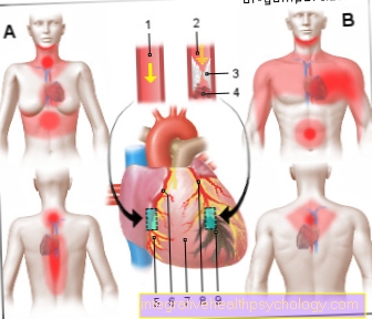
A systemic complication can be heart failure. The short circuit between artery and vein leads to an increased cardiac output and, as a result, increased stress on the heart. Another complication is the so-called steal phenomenon. This leads to circulatory disorders in the area below the shunt, as the short circuit virtually “steals” blood. A steal phenomenon is manifested by a cold hand accompanied by pain or numbness.
Clogged dialysis shunt
Frequent punctures of the dialysis shunt lead to changes in the vessel wall. These include above all constrictions (Stenoses) through tissue growth or the formation of bulges in the vessel wall (Aneurysm). These changes reduce the flow of blood through the shunt and can lead to a complete occlusion of a blood clot (thrombosis) come. In this case, you have to react quickly to enable recanalization. An operation is necessary on the same day. A shunt occlusion can be detected by the patient because there is no normally audible buzzing over the shunt.
If there is a shunt occlusion, the thrombus must be removed with a catheter intervention or open surgery. As part of the operation, it should also be checked why there was a shunt occlusion and the causes eliminated.
In rare cases, the dialysis shunt cannot be reopened and a new shunt must be placed.
Bleeding from the shunt
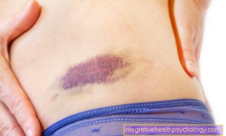
An incorrect puncture of the dialysis shunt can lead to bleeding. However, this bleeding is usually small and has no further consequences for the patient. This can lead to the formation of a bruise (Hematoma) come. If the bleeding is larger than expected, surgery may be necessary in rare cases to ensure the function of the shunt and to find out the exact cause of the bleeding.
However, with regular blood clotting checks and a careful puncture, the risk of bleeding from the shunt is very low.
Where can you place a shunt everywhere?
In principle, the dialysis shunt should be placed on the non-dominant extremity. In the case of right-handers, the system should be placed on the left arm and vice versa. As a result, the patient is not so restricted in his everyday movement.
In most cases, the dialysis shunt is placed on the upper extremity. The most common shunt connection here is the so-called Cimino shunt. It lies on the forearm and connects the radial artery and the cephalic vein. Another option is to connect the brachial artery and the cephalic vein in the crook of the elbow. If it is not possible to apply a shunt to the arm, in rare cases it may also be possible to apply a shunt to the thigh.
Further information
More information on this topic:
- dialysis
- Kidney failure
- Kidney transplant
- Heart failure
You can find an overview of all topics in the field of internal medicine under: Internal medicine A-Z


.jpg)