The anatomy of the arm
General
The human arm, also known as the free upper extremity, is the transformation or further development of the front limb into a gripping tool.
However, it not only serves as a gripping tool, but also for balancing when walking upright.

Function of the arm
The upper limb of man has the greatest possible freedom of movement all parts of the body. This is due to the mobility in
- shoulder-
- Elbow- and
- wrist enables.
anatomy
The arm is divided into the
- upper arm,
- forearm and the
- hand.
The individual parts of the arm protrude Joints in connection. In addition to connecting the individual parts of the arm, these are also used to perform numerous movements.
Of the upper arm consists of a large tubular bone (Humerus). This one is about that Shoulder joint with the shoulder and thus connected to the body trunk. This is one Ball joint, which allows three different directions of movement.
Of the upper arm can on the one hand be a sagittal axis be moved.
This is done by pulling the arm towards the body from the side (Adduction), as well as the lateral movement of the arm away from the body (Abduction). The abduction limit is 90 °, the movement over 90 ° is called a lifting movement (elevation) designated. There are different for these movements Muscle groups responsible that as
- Adductors,
- Abductors, or.
- Elevators are designated.
Furthermore, a movement of the arm is one frontal axis possible. This means lifting the arm forward (Anteversion), or the return of the arm (Retro version). The last option is to rotate the arm in the Shoulder joint.
This rotation can be inward (Internal rotation) or outwards (External rotation) are executed. The rotation in the shoulder joint serves to support the rotation in the Forearm:
- Turning the palm up = Supination,
- Rotation of the palm down = pronation.
The Elbow joint serves as the connection between the humerus (Humerus) and the two forearm bones (Cubit and spoke). Due to the various extensor and flexor muscles, a Elongation and diffraction take place in the elbow joint.
But also the one named above Pronation and Supination is only possible due to the rotation of the spoke head in the elbow joint.
in the wrist, on the one hand, articulate the body-hugging (proximal) Carpal bones with the bones of the forearm and the rows of carpal bones one below the other. These different joints make that possible
- Diffraction (Flexion, Plamarflexion) and
- Elongation (Extension, Dorsiflexion),
- as well as a Splaying movement of Wrist. This can be done in the direction of the thumb (Ulnar abduction), as well as in the direction of the little finger (Radial abduction) are executed.
The finger themselves consist of numerous small bones, which are individually connected by numerous joints, so that the fingers
- bent and
- stretched can be.
- Furthermore, all fingers can be moved away (Abduction) and
- introduced (Adduction) become.
Of the thumb can be the only finger left Opposition movement To run. The thumb is moved to the palm of the hand. In addition to the bony connections between the various parts of the arm, the Muscles to connect the individual structures. In addition to the connection, they also convey the individual movements of the three essential joints and serve the Power transmission.
Because of this, the individual pull Muscles always over one of the various joints in order to get it moving. Even the individual Vessels and annoy arise on the shoulder or the trunk of the body and then continue down to the individual fingers. So the supply takes place through arterial blood and the nerval Innervation.
The Veins and Lymphatic vessels however, collect the blood in the Periphery, so the fingers and then feed this to the trunk of the body. Thus the veins and lymph vessels of the arm are connected to each other or merge into each other and thereby transport the different ones liquids.
Upper Arm Anatomy
The upper arm is part of the upper extremity and consists of a bone, several muscles, and other structures.
The upper arm is connected to the trunk via the shoulder joint. This joint makes the entire arm very flexible. Towards the forearm, the upper arm closes with the elbow joint.
The only bone on the upper arm is the humerus bone (Humerus). This large tubular bone, together with the socket of the shoulder blade, forms the shoulder joint. This joint is stabilized by a capsule and several muscles. This muscle group is known as the rotator cuff, because it encloses the shoulder joint like a cuff and the muscles are responsible, among other things, for the rotational movement (rotation).
These muscles belong
- the teres minor muscle, the subscapularis muscle,
- the muscle supra and
- infraspinatus.
Another important muscle that attaches to the upper arm is the biceps (Musculus biceps brachii). This muscle has several functions and is responsible for movement in both the shoulder and the elbow joint. The arm can be turned inward, stretched forward, moved away from the body and bent at the elbow. The brachialis muscle is also responsible for flexing the arm. On the back of the upper arm there is the triceps muscle (musculus triceps brachii). This stretches the arm in the elbow joint and is able to pull the arm back towards the body.
The blood supply to the upper arm is ensured by the brachial artery, which in turn is divided into several branches. The venous outflow is accomplished through several veins, such as the superficial basilica and cephalic veins. The two nerves, the musculocutaneous nerve and the radial nerve, innervate the muscles in the upper arm and sensitive areas of the skin.
Learn more at: Upper arm muscles
Forearm anatomy
The forearm, like the upper arm, belongs to the upper extremity. It is connected to the hand via the wrist and to the upper arm via the elbow joint. In contrast to the upper arm, two bones form the basis of the forearm, the ulna and the radius.
These two long bones are connected by a membrane, the membrana interossea antebrachii. In addition, these bones together form a joint on the elbow and wrist, the proximal and distal radioulnar joint. The main movements that arise from this joint are pronation and supination of the forearm and wrist, respectively.
The muscles of the forearm consist of many muscles that can be functionally divided into the flexor muscles and the extensor muscles. The muscles that bend the hand can be divided into the deep and superficial muscles.
The deep muscles include the flexor digitorum profundus muscle and the flexor pollicis longus muscle. The superficial flexors include a total of five muscles, for example the pronator teres muscle.
The extensor muscles also have superficial and deep muscles. There is also another muscle group, the radial group. These muscles are responsible for kinking the hand towards the radius.
The blood supply takes place through the arteria ulnaris and arteria radialis. These two vessels arise from the brachial artery. The numerous muscles are supplied by several nerves, such as the radial and ulnar nerves.
Find out more at: Forearm muscles
Hand anatomy
The hand is a complex structure with many bones and muscles that allow great mobility. Their function is to grasp and hold objects, without which an independent life is not possible. The hands are connected to the forearm via the wrist and thus form the last part of the upper extremity.
The hand consists of a total of 27 bones, which makes up around a quarter of all human bones.
There are eight carpal bones (scaphoid, moonbone, triangular bones, pea bones, large and small polygonal bones, headbones, hooked bones), five metacarpal bones and 14 finger bones. The fingers are made up of three small bones. An exception is the thumb, which consists of only two bones.
In addition to the many bones, there are 33 muscles that are involved in the great mobility. Most of them have their origin in the forearm and pull into the hand with their tendons.
The blood supply to the hand is ensured by the arteria radialis and the arteria ulnaris.
The motor and sensitive supply of the hand is also taken over by several nerves (radial nerve, ulnar nerve and median nerve). Depending on which nerve is injured, there are characteristic deficits in the hand, such as a falling hand. This gives an indication of damage to the radial nerve, which can be injured, for example, by a fracture of the upper arm.
Find out more at: Muscles of the hand
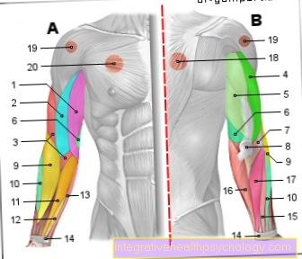
Illustration of the arm muscles

Arm muscles
- Two-headed upper arm muscle
(Biceps) short head -
M. biceps brachii, caput breve - Two-headed upper arm muscle
(Biceps) long head -
M. biceps brachii, caput longum - Upper arm muscle (arm flexor) -
Brachialis muscle - Three-headed upper arm muscle
(Triceps) side head -
M. triceps brachii, caput laterale - Three-headed upper arm muscle
(Triceps) long head -
M. triceps brachii, Caput longum - Three-headed upper arm muscle
(Triceps) inner head -
Triceps brachii muscle,
Caput mediale - Knobby Muscle - Muscle anconeus
- Elbow - Olecranon
- Upper arm spoke muscle -
Brachioradialis muscle - Long spoke-side hand straightener -
Muscle extensor carpi radialis longus - Spoke-sided hand flexor -
Muscle flexor carpi radialis - Superficial finger flexor -
Muscle flexor digitorum superficialis - Long palm tendon tensioner -
Palmaris longus muscle - Extensor tendon strap -
Retinaculum musculorum extensorum - Short spoke-side hand straightener -
Muscle extensor carpi radialis brevis - Elbow-sided hand flexor -
Muscle flexor carpi ulnaris - Finger extensor -
Muscle extensor digitorum - Trapezius -
Trapezius muscle - Deltoid -
Deltoid muscle - Pectoralis major -
Pectoralis major muscle
You can find an overview of all Dr-Gumpert images at: medical illustrations
Diseases of the arm
Poor falls asleep - what can be the cause?
There are several causes that can lead to an arm falling asleep. These are usually harmless, but there are also serious illnesses that manifest themselves with such complaints. An arm that has fallen asleep leads to tingling or numbness in the affected arm, which can be perceived as very uncomfortable. Sometimes pain and mobility restrictions can occur.
The most common cause is poor arm posture, so that a nerve is pinched off. First, the sensitivity is disturbed, which is transmitted to our brain as tingling or numbness. It helps if you move your arm so that the clamped nerve is relieved. Usually the feeling in the arm normalizes within a short time and there is no damage left.
If the arms or other parts of the body regularly fall asleep even though the posture is not uncomfortable, a doctor should be consulted. In rare cases such symptoms hide serious illnesses that require therapy.
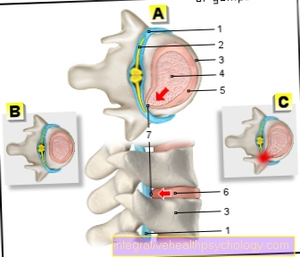
These include, among other things, slipped discs or the nerve disease multiple sclerosis. People with diabetes (diabetics) must also pay particular attention. This metabolic disorder can also damage nerves in the long term, which can lead to polyneuropathy. In the case of polyneuropathy, sensitivity is lost, especially in the feet, and patients often feel tingling or numbness. This disease can be counteracted by a good control of the diabetes.
Read on under:
- Hand falls asleep at night
- Circulatory disorders in the arm
Broken bones on the arm
Fractures on the arm occur in all age groups and are among the most common bone fractures. In principle, any bone on the arm can be broken.
The most common break on the arm is the distal radius fracture. In most cases, this injury is caused by falling on an outstretched arm. The spoke (radius) is broken and, depending on the extent, surrounding structures can also be damaged.
Another common fracture on the arm is the humeral head fracture. This injury particularly affects the elderly over the age of 70 and postmenopausal women. In this fracture, the fracture gap is in the upper area of the upper arm bone (humerus). This fracture is usually the result of a fall, but there are also less common causes such as bone metastases.
The symptoms of fractures in the arm are independent of the fracture site and usually consist of pain, swelling, redness and restricted mobility. The diagnosis of a fracture can often be made immediately after the clinical examination, but this is confirmed by an X-ray. The X-ray images are taken from two planes in order to be able to detect possible bone displacement.
In the case of open or complicated bone fractures, an operation is performed, otherwise a simple plaster cast is sufficient, which usually has to be worn for six to eight weeks.
Learn more at:
- Broken upper arm - you need to know that now!
- Broken spoke
- Radial head fracture
Arm pain - what's behind it?
Pain in the arm can have numerous causes and is usually harmless. In the case of the pain history, particularly sporting activities or falls should be inquired about. Often there are muscular causes behind pain in the arm area, which arise, for example, from overexertion during exercise or tension. Over-stimulation of the muscles or nerves should be considered, especially in competitive athletes, such as tennis players. In general, a muscle strain, muscle contusion, a sprain of a joint or a broken bone should always be excluded.
In older patients, previous falls should be clarified, as falls from a small height can damage bones. Reduced bone density (osteoporosis), which older women in particular suffer from, are beneficial for such injuries.
For arm pain, inflammation can also be an issue, often in the area of the joints. In these areas, the tendons of the muscles and also the bursae are often irritated, which can lead to severe pain. Chronic inflammatory diseases of the joints such as rheumatism are also expressed as pain.
Of course, there are other more serious conditions that can cause arm pain. However, these are less common and often manifest through other symptoms. A mass in the area under the armpit, for example due to swollen lymph nodes as part of an infection or a malignant process, can radiate painfully into the arm.
Further details can be found at:
- Left arm pain
- Pain in the right arm
- Pain in the outer upper arm
- Pain in the forearm
Dislocated arm
A dislocated arm is the most common dislocation (dislocation) of a joint in Germany. In most cases, the arm dislocates after a traumatic event, usually after falling onto the outstretched arm.
The most common dislocation of the shoulder is what is known as an anterior shoulder dislocation. The head of the humerus is no longer in its socket, but is shifted forward and down. Typical symptoms of this injury are shoulder pain, restricted mobility and a resilient position of the humerus. In addition, the empty socket and the dislocated head of the humerus can usually be felt.
A dislocated arm is confirmed by an X-ray from two planes. Two recordings from different perspectives are always necessary in order not to be able to detect a dislocation to the front or to the rear.
The treatment consists of an immediate manual reduction of the shoulder joint, during which the patient is usually sedated and additionally given pain medication. If this reduction is successful, the pain will subside very quickly and the affected arm should be immobilized with a bandage. In serious cases, for example with injured vessels or nerves or with repeated dislocation of the shoulder, the joint should be treated surgically.
Learn more about this topic: shoulder dislocated
Sprained Arm - What Can I Do?
A sprain describes a severe overstretching of ligaments or joint capsules and can affect different parts of the body. The foot and knee joints are particularly at risk, but you can also sprain your arm due to excessive strain. A sprain or distortion is characterized by pain and, in most cases, swelling. The swelling is caused by a bruise.
In the case of uncomplicated sprains, you do not necessarily need to consult a doctor. The so-called PECH scheme gives a good overview of the treatment. The four letters stand for the four important pillars that support rapid regeneration of the injured joint: rest, ice, compression and elevation. After a sprain, physical activity, especially sport, should be avoided for a few weeks. In addition, the injured area should be cooled as quickly as possible and a bandage applied with light pressure. To further relieve the arm, it should be raised.
All of these measures will help prevent an effusion. Usually the pain subsides after a few weeks and you can slowly go back to your sporting activities. If the pain persists or is very severe, however, a doctor should be consulted who can rule out a possible torn ligament or something similar.
Find out more at:
- Hand sprain
- Sprained thumb
Arm is shaking
A tremor or tremor of the arm can have a variety of causes, which are usually completely harmless. Despite everything, you should observe increased tremors, which can also affect a leg, for example, and have it checked out by a doctor.
A minimal tremor of the muscles, which we normally do not notice, is normal and has no disease value. However, if these muscle twitches intensify, one should pay attention to the situations in which this occurs. There are many types of tremors, for example that during physical rest (resting tremor) or during active movements (movement tremor).
A common cause that can lead to increased muscle activity is severe psychological stress. However, medications such as antidepressants or heavy caffeine or nicotine consumption can also cause the arm to tremble.
If these causes can all be ruled out, neurological causes in particular must be considered. A pathological change in the nerves or the brain can be noticeable, among other things, by a tremor of the extremities. Well-known examples are multiple sclerosis (MS) and Parkinson's disease. These two neurological diseases each have a characteristic tremor as a symptom.
The patients with multiple sclerosis show a so-called intention tremor. This tremor mainly occurs when patients begin specific movement. In contrast, Parkinson's patients tend to have a postural tremor, which is most pronounced when the body is at rest.
Read also under:
- Hands are shaking
- Muscle twitching in the upper arm
Arm is swollen on one side
A one-sided swollen arm is always a reason to consult a doctor, especially if the swelling occurs suddenly and for no explainable cause. In general lymphatic disorders, which can also be congenital, usually both arms are affected.
One-sided swelling of the arm often affects women who have had to undergo surgery because of breast cancer. During this operation, not only the tumor tissue in the diseased breast is removed, but also affected lymph nodes, depending on the extent of the tumor. These are located in the armpit area and are important for lymph drainage. If these lymph nodes are missing, the lymphatic drainage is disrupted, causing the arm to swell. This can sometimes lead to pain, so that the range of motion is limited.
The treatment of such lymph drainage disorders consists of several components. On the one hand, sufficient exercise and healthy skin care have a positive effect on the development of such diseases. Manual lymph drainage has proven to be particularly effective. Trained physiotherapists stimulate the lymphatic drainage with certain movements and massages so that the congestion is released. Compression bandages or stockings, which compress the arm and thus promote lymphatic drainage, also have a supportive effect.
Another cause of unilateral arm swelling is an insect bite. This is very rare but should be examined by a doctor and treated if necessary.
You can also find out more at: Lymphatic system
Kinesio tape on the arm
A kinesio tape is one of the alternative healing methods and is used, among other things, for arm diseases. The kinesio tape is an elastic cotton tape with an acrylic adhesive layer that is applied to the skin. How to properly tape the arm depends on the injury.
The kinesio tape is helpful, for example, with a so-called tennis elbow or tendonitis. However, there are general guidelines that should always be followed when applying kinesio tape.
Before applying the tape, the skin must be depilated and cleaned with soap. In order for the tape to stick longer, it helps to round off the corner and not to touch the adhesive surface while sticking it on. Before sticking the tape on, you should warm it up slightly, for example by rubbing it briefly between your hands.
The kinesio tape is supposed to relieve muscles and joints and there are numerous different ways of taping. With tennis elbow, the arm is physically overloaded and the pain is on the outside of the elbow. The arm is extended at the elbow joint and flexed at the wrist while taping. The first longer kinesio tape is attached to the outside of the elbow, starting from the back of the hand over the forearm. The second tape is shorter and is pulled under tension from the lower inner elbow to the back of the elbow. The right pull at the optimal point is crucial. To ensure this, it is recommended that such tapes only be attached by experienced therapists.
Further information can be found at: Tennis elbow- you should know that!
Summary
The human arm with its anatomical structure is a human organ with the greatest possible range of motion.
In addition to the individual movements, it also serves to attitude of Equilibrium. For this reason, people swing back and forth with their arms when walking. The numerous Movements and Functions are through
- the three great bony parts of the arm,
- their articulations and
- Muscles.
The blood- / Lymphatic vessels and annoy of the arm usually run in a connection with the shoulder up to the fingers and thus supply the entire arm Nutrients and oxygen.
From the large communicating vessels numerous branch smaller ones Vessels in order to be able to treat the entire arm. The individual nerves are also responsible for different structures of the arm, so that the injury of a nerve on the upper arm also leads to the failure of individual muscles on the forearm or the hand can lead.