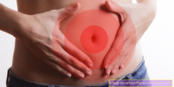
The breathing
Synonyms
Lungs, airways, oxygen exchange, pneumonia, bronchial asthma
English: breathing
definition
Breathing is needed to supply the body with oxygen.
To do this, the body absorbs oxygen from the air via the lungs (Pulmo) and releases it again in the form of carbon dioxide (CO2).
The regulation of breathing is subject to complex control mechanisms and is achieved by many different muscle groups.
Respiratory chain
The respiratory chain is a vital process that takes place in the mitochondria. This is basically about energy generation. So-called reduction equivalents (NADH + H + and FADH2) are formed from components of our food such as sugar, fat and protein before the respiratory chain. These reduction equivalents are then used in the respiratory chain via various complexes to produce ATP (adenosine triphosphate).
The respiratory chain consists of 5 complexes, which are located in the inner mitochondrial membrane. In simple terms, a proton gradient is built up over the first 4 complexes. This means that there are many protons outside the membrane and thus an imbalance arises. To compensate for this imbalance, the direction of flow is directed towards the interior of the membrane. The 5th complex of the respiratory chain makes use of this pressure and uses the flow of protons to produce ATP.
ATP is a universal supplier of energy and is required everywhere in our body (for example for muscle activity or chemical processes in cells). In total, 32 ATP can be produced from one sugar molecule, which can then be used. If the respiratory chain is no longer active, this has serious consequences. So-called cyanides, also known as hydrogen cyanide, inhibit the respiratory chain and thereby prevent the formation of ATP. This leads to death within a short time.
You might also be interested in this topic: Cellular respiration in humans
Respiratory muscles
The muscles that work for the inflow of air into and out of the lungs are called respiratory muscles.
The most important respiratory muscle is the diaphragm. It is a quasi-ring-shaped, flat muscle that forms the border between the thoracic and abdominal viscera and is attached to the edge of the abdominal wall and the spine.
When the diaphragm is relaxed, the middle part arches like a hemisphere into the chest, as there is less pressure here than in the abdomen. If the musculature is now tense, the diaphragm sinks and becomes almost horizontal. This increases the volume in the thorax (chest) and thus in the lungs.
This means that the pressure in the lungs is lower than in the air. This negative pressure represents the driving force for the influx of air (inhalation, inspiration). Parts of the intercostal muscles and individual muscles of the shoulder girdle can, depending on the posture, support inhalation (auxiliary breathing muscles).
Read more on the topic:
- Diaphragmatic breathing
- Abdominal breathing
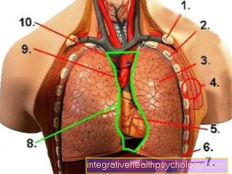
Illustration of the lungs
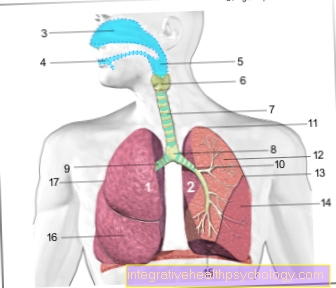
- Right lung -
Pulmodexter - Left lung -
Pulmo sinister - Nasal cavity - Cavitas nasi
- Oral cavity - Cavitas oris
- Throat - Pharynx
- Larynx - larynx
- Windpipe (approx. 20 cm) - Trachea
- Bifurcation of the windpipe -
Bifurcatio tracheae - Right main bronchus -
Bronchus principalis dexter - Left main bronchus -
Bronchus principalis sinister - Lung tip - Apex pulmonis
- Upper lobe - Superior lobe
- Inclined lung cleft -
Fissura obliqua - Lower lobe -
Inferior lobe - Lower edge of the lung -
Margo inferior - Middle lobe -
Lobe medius
(only on the right lung) - Horizontal cleft lung
(between upper and middle lobes on the right) -
Horizontal fissure
You can find an overview of all Dr-Gumpert images at: medical illustrations
Activation of the auxiliary respiratory muscles
Everyone knows the image of an exhausted athlete who activates his auxiliary breathing muscles by leaning forward and supporting his upper body with his hands on his thighs. This gives the auxiliary breathing muscles a more favorable leverage ratio and can ventilate the lungs well, saving effort.
If inhalation is done through active work, it would make sense if the body used the energy provided for exhalation.
And that's exactly what the body does, at least at rest. The diaphragm relaxes and returns to its resting position with the curvature in the chest cavity. This increases the pressure there and the air is pressed out of the lungs. As the breathing rate increases, the time to exhale must decrease. Then the body uses its exhalation muscles. Parts of the intercostal muscles, but also the abdominal muscles, are crucial here.
You may also be interested in this topic: Chest breathing
All the muscles of breathing
Inhalation muscles (inspiratory muscles)
- Diaphragm = most important respiratory muscle
- Musculi intercostales externi (external intercostal muscles)
- Levatores costarum muscles (rib lifter)
- Scalen muscles
- Serratus posterior superior muscle
- Serratus anterior muscle (anterior saw muscle)
- Rectus abdominis muscle (straight abdominal muscle)
Exhalation muscles (expiratory muscles)
- Musculi intercostales interni et intimi (inner intercostal muscles)
- Abdominal muscles
- Serratus posterior inferior muscle
- Retractor costae muscle
- Transversus thoracis muscle
- Subcostal muscle
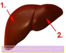
Structure of the thorax
- Collarbone
- rib
- lung
- Chest wall
- heart
- diaphragm
- liver
- Mediastinum
- Skin artery (aorta)
- Superior vena cava (Vena cava)
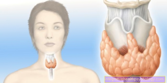
Bronchial muscles
The Bronchial muscles has a kind of control function for the distribution of the breathing air to the individual sections. It is usually arranged in a spiral around the airways and is particularly numerous in the small and medium-sized ones Bronchi.
This makes sense, since the walls have less cartilage with increasing distance from the neck and can therefore be changed significantly more in diameter through contractions. In the bronchi, which are supposed to get a lot of air, the muscles relax and the diameter of the bronchi widens. In the opposite case, tensing the muscles ensures a reduced diameter and thus less ventilation of the lung section.
The bronchial muscles play a larger, if not necessarily wanted, role Exhale. If the muscles are tense and the diameter of the bronchius is narrow, it may be that not enough air can flow out of the alveoli during the exhalation phase. Now, during the next inhalation, more air comes in that cannot flow out sufficiently during the next breath. This mechanism will obstructive (= occlusive) pulmonary dysfunction called. In the long run, the affected alveoli literally slacken - in this case one speaks of a Emphysema.
Now, of course, you can ask yourself why more air comes in when you breathe in than can come out when you breathe out. The reason is as follows: When inhaling, there is negative pressure in the lungs, which of course also has an expanding effect on the bronchi. Exhalation is triggered by overpressure in the lungs - this overpressure also compresses the airways.
The bronchial muscles are of the so-called type smooth muscles. This means that it works without conscious control but receives its impulses from it vegetative (autonomic) nervous system.
The two parts of the autonomic nervous system (sympathetic nervous system (short: sympathetic) - parasympathetic nervous system (short: Parasympathetic nervous system)) have a nonsensical effect.
As with all connections between nerves and muscles, the respective effect on the muscle is mediated by proteins of the cell membrane (receptors), which can convert the signal of the nerves into muscle excitation or relaxation via a change in shape.
During stress and physical work, the sympathetic nervous system a signal for relaxation of the bronchial muscles and thus for a widening of the airways (bronchodilation). This is mediated via so-called beta-2 receptors, which are located on the cell membrane of the muscle cells.
In the case of shortness of breath (dyspnea), caused by increased tension in the bronchial muscles, special medications (beta-2 sympathomimetics) are given that alleviate the symptoms as they mimic the effect of the sympathetic nervous system on the receptors (mimetic = imitate) .
Of the Parasympathetic nervous system, which is active during rest and sleep, leads to tension in the muscles and thus to a narrowing of the airways (bronchoconstriction).
There are other substances that can cause the bronchial muscles to contract, the most important being histamine. This histamine is released by special defense cells (so-called mast cells) as part of an allergic reaction. The amount of histamine is usually so large that the muscles cramp. This makes breathing of the patient life-threateningly difficult. This condition is known as an asthmatic attack (asthma attack).
Difference in breathing in adults and babies
Breathing in a baby and an adult is different in certain ways. But the mechanism of breathing is the same. Inside the womb, the baby's lungs are filled with fluid. The mother's oxygen-rich blood supplies the baby at the time.
From birth, the baby breathes like adults through the expansion and contraction of the lungs. The frequency of breathing is increased in babies compared to adults. While an adult human breathes around 12-15 breaths per minute, a newborn breathes around 40 times per minute.
In an infant, about 30 breaths per minute can be determined. This may seem like a lot at first and may scare some parents, but rapid breathing is perfectly normal. Breathing sounds are also a concern. While adults hardly make any breathing noises and a whistling or rattling can usually be heard when they are ill, babies can often hear noises breathing.
This is due to the fact that the baby's mucus is difficult to carry away and remove. Adults, for example, blow their noses more often, while in babies the mucus remains in the nose and can thus lead to noises. Other than that, there are no differences in breathing.
This article might also interest you: Bronchitis in the baby
Breathing techniques for specific situations
Breathing when you go into labor
The onset of labor heralds the imminent birth. As the contractions progress, the intervals become smaller and smaller. At this point it is still important to keep to a certain breathing pattern. In this case, it is advisable to inhale deeply into your stomach at the beginning of a contraction and then slowly let the air out again.
It is often helpful for women who are signing to make certain sounds such as “Aaah”, “Uhhh” or “Ohh” in order to support the slow, controlled exhalation of the air. It is also advised to breathe in through your nose and breathe out through your mouth.
Find out more about the topic: The different types of labor
Breathing at birth
In the transitional phase of childbirth, i.e. when pressure can be felt on the pelvic floor after the onset of labor, no pressure should be generated to force the baby out. For this reason, “panting” is recommended during the transitional phase of labor. Here you exhale in many small breaths.
During the expulsion phase of the birth, active pressing should be done. In most cases, you inhale deeply before pressing and then exhale again after pressing. However, it is important not to hold your breath for too long in order to maintain the oxygen supply; on the other hand, it is also important not to breathe too quickly, as this can lead to hyperventilation and circulatory problems. In most cases, however, breathing works very well intuitively or with guidance. The tips and exercises in antenatal classes can also help many women with childbirth.
Read more on the topic:
- Breathing at birth
- Breathing exercises
Breathing while jogging
Breathing while jogging is a topic that has been discussed in the sports world. In the past, people were advised to keep a strict breathing rhythm (about 2 steps for inhalation, 3 steps for exhalation). Nowadays it is believed that a steady rhythm tends to limit runners and lead to problems. Abdominal breathing is now mainly recommended. Abdominal breathing is driven by the diaphragm, which contracts and thus expands the entire lungs.
Read more on the topic: Abdominal breathing
By contrast, chest breathing mainly unfolds the upper part of the lungs. As a result, the volume of the lungs is not used adequately. It is even recommended to practice abdominal breathing outside of jogging, for example with yoga. Apart from that, it is recommended that you breathe through both your nose and mouth. Nasal breathing has the advantage that the air is warmed and moistened via the mucous membranes of the nose. However, the breathing volume is restricted by the small diameter of the airways in the nose. When breathing through the mouth, a higher volume of breathing can be achieved, but a dry throat is also more common.
Find out more about the topic: Stitch
Breathing while crawling
The crawl is a special swimming technique in which the swimmer has his head under water and turns his face to the surface of the water to breathe. The act of breathing should take place in the shortest possible time, as the head has a higher resistance above water and thus makes the swimmer slower. The head collapses sideways and the swimmer inhales. When it comes to speed, breathing is usually done through the mouth, as breathing through the mouth allows a larger volume of air to be breathed in less time.
Read more on the topic: Freestyle swimming
However, if you swim long distances, the mouth and throat area can quickly become dry. In this case, it is better to inhale through the nose. Exhaling while crawling takes place underwater. It is not necessary to lift your head above the surface of the water and would mean an unnecessary loss of time.
Breathing in fear
Everyone has felt fear at some point. The heart starts racing and the chest feels constricted. Breathing also becomes faster and shallower. Sometimes you even hold your breath out of fear. However, there are also breathing exercises that help against anxiety. By using the breathing techniques, one begins to relax and does not let fear have such great control over the body. First of all, it is important to consciously breathe more slowly. An adult person breathes around 12 to 15 times per minute, usually more often in a fearful situation.
You should try to get a frequency of about 6 breaths per minute. This goes hand in hand with breathing in and out very slowly and deeply. After exhaling, you can also take a short break until you feel the urge to inhale again. To slow down the exhalation, it is helpful to exhale through the slightly closed lips and thereby slow down the air. A long exhalation is particularly helpful in regulating your breathing and being able to relax.
Read more on the topic: Breathing exercises to relax
Ideal breathing for falling asleep
For some time now, the so-called 4-7-8 breathing technique has become very popular as a sleep aid. It is a special breathing technique developed by the American doctor Dr. Andrew Weil was developed. It is based on breathing exercises from yoga and is said to have a very relaxing effect, so that you can fall asleep within a short time. The advantages of this exercise are that it is free, unsupervised, and takes less than a minute.
First you breathe in through your nose for four seconds. Then the breath should be held for 7 seconds. Finally, the air should be exhaled again within 8 seconds, while the tip of the tongue is placed on the roof of the mouth, i.e. behind the upper incisors. This exercise lowers the pulse and relaxes you. This makes it easier for many people to fall asleep quickly. Alternatively, there are other exercises to help you fall asleep quickly. The basic idea is always that you concentrate on your breathing and breathe consciously.
On the one hand, this forces you to ignore your thoughts and worries that keep you from sleeping. In addition, conscious, calm breathing has a relaxing effect. For example, you can place your hands on your chest or stomach and deliberately inhale slowly from top to bottom. Breathing should flow like a wave from top to bottom. Then you let the air out again from bottom to top. It is important to feel the movement of the breath with your hands and to focus on it.
Read more on the topic:
- Breathing exercises to help you fall asleep
- Difficulty falling asleep
Lung disease with difficulty breathing
asthma
There are different forms of asthma (bronchial asthma). The most common form is allergic asthma. Here, an allergy-causing irritant (allergen) leads to a histamine (see above) mediated constriction of the lung branches (bronchi). It is characteristic that the inhaled air can no longer leave the lungs. A characteristic sign of the disease is shortness of breath.
Further information is available under our topic: asthma
lung infection
Inflammation of the lungs (pneumonia) is mostly caused by bacteria. Inflammatory infiltrates (immune cells and bacteria) lead to the filling of the alveoli, which are then no longer available for gas exchange.
Characteristic symptoms are:
- fever
- to cough
- shortness of breath
Further information is available under our topic: Signs of pneumonia
COPD
The chronic obstructive pulmonary disease (disease) is caused in particular by smoking. In particular, breathing in the air is difficult due to the permanent constriction of the bronchi. Their characteristic symptoms are shortness of breath, expectoration, and coughing.
Read more about this: COPD
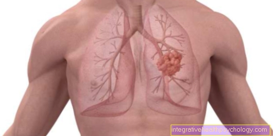
Lung cancer
Lung cancer is also mainly caused by smoking and in most cases leads to death of the patient.
There are no typical symptoms that are unique to lung cancer.
You might also be interested in this topic: How do you recognize lung cancer?
Recommendations from the editorial team
- Human breathing
- Respiratory muscles
- Diseases of the lungs
- Shortness of breath
- asthma