Overview of human eye diseases
There are a variety of diseases in the eye, which often have many different causes. Inflammation, injury, and change in old age can transform and damage the eye. Here you will find a selection of the most common diseases of the eye.
The most common eye diseases are listed below, sorted by:
- Eye diseases that often occur in old age
- Inflammation and infection in and around the eye
- Eye diseases as a result of other underlying diseases
- Tumors and abnormalities in and around the eye
- Eye disorders due to nerve damage
- Vision disorders
- Misalignments of the eyes
Eye diseases that are common in old age

Glaucoma / glaucoma
Glaucoma (glaucoma) is less of a specific disease, but more of a generic term for a series of diseases that are associated with typical damage to the optic nerve papilla and the visual field. This can lead to damage to the optic nerve head and the retina, resulting in visual field defects and, in extreme cases, blindness of the eye. The therapy depends on the cause and the underlying disease. However, in the event of discomfort to the eye, an ophthalmologist should be consulted for further clarification.
Further information on this topic is available at: Green Star
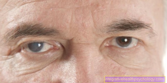
Cataracts
The cataract generally describes any form of lens opacity. In an advanced cataract, a gray haze can be seen behind the pupil.
The clouding makes the lens impermeable to light and there is a gradual decrease in vision up to complete blindness and a deterioration in visual acuity. The main symptom of cataracts is deteriorating eyesight. The causes are very diverse. If the cataract-induced lens opacification deteriorates significantly and normal vision is severely impaired, surgery is the only treatment option.
You can find more information under our topic: Cataract
Age-related macular degeneration (AMD)
A distinction is made between a dry age-related macular degeneration (85%) and a wet form (15%). The causes of AMD are not yet fully understood. With macular degeneration, those affected usually notice gray shadows in the central field of vision, i.e. exactly where they are looking. Visual acuity is very impaired, often so bad that it is hardly possible to read. There is no established therapy for the dry form of age-related macular degeneration.
Better treatment options are known for the wet form, such as laser surgery and surgical rotation. Magnifying visual aids (magnifying glasses, magnifying glasses, screen readers) are used to alleviate the symptoms that are expressed in the increasing deterioration of visual performance.
Further information on the subject can be found at: Macular degeneration
Inflammation and infection in and around the eye
Barley grain (hordeolum)
The stye is a purulent inflammation of the eyelid glands. It can occur on the inside of the eyelid as inflammation of sebum glands (the so-called meibomian glands) or on the outside as inflammation of sweat glands (Moll glands) or sebum glands (Zeis glands).
The main symptom is a painful lump on the edge of the eyelid. The treatment is carried out with antibiotic ointment and heat treatment by means of red light radiation.
Further information is available under our topic: Stye
Hailstone (chalazion)
Hailstone is an inflammation of the sebum glands on the inside of the eyelid (so-called meibomian glands) due to the accumulation of secretions. Unlike the stye, it is not painful. The impairments are mostly of a purely cosmetic nature:
The hailstone is visible as a nodular swelling of the eyelid, which can reach a considerable size. The treatment is carried out surgically by puncturing and clearing out the secretion mass.
Further information is available under our topic: Hailstone
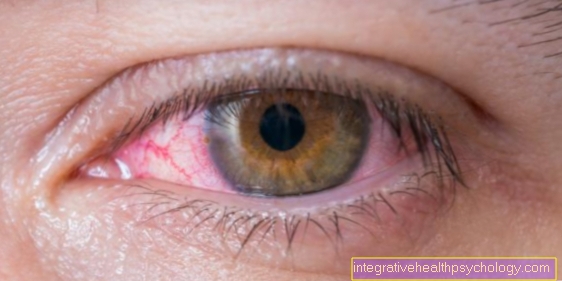
Conjunctivitis
Inflammation of the conjunctiva is one of the most common diseases of the eye. The eye itches, is red and gives off secretions. It can be triggered among other things by bacteria, viruses, an allergy or external stimuli such as dry air. Depending on the cause, it may or may not be contagious. The main symptoms are often a red eye, swelling, secretion and sometimes pain. Since conjunctivitis (Conjunctivitis) can be triggered by several causes, there are also different treatment approaches. You should be careful with self-treatment without consulting a doctor.
Further information on this topic can be found at: Conjunctivitis
Trachoma
Trachoma is a chronic conjunctivitis caused by the bacterium Chlamydia trachomatis (Conjunctivitis), which often leads to blindness. It usually manifests itself within 5-7 days with weeping conjunctivitis with a foreign body sensation. Systemic or local, intracellularly effective antibiotics are used to treat trachoma. A doctor should be consulted for further clarification.
Further information on this topic can be found at: Trachoma
Ocular herpes
The ocular herpes describes an infection of the eye with herpes viruses. Different structures of the eye can be affected (nerves, cornea, etc.). In addition to ocular herpes, the herpes simplex viruses also often cause herpes simplex keratitis, i.e., herpes-related corneal inflammation. The main symptoms are often reddening of the eyes, a foreign body sensation, and severe burning and itching. The treatment is carried out using eye ointments and / or eye drops. However, a doctor should be consulted before treatment.
For more information, see: Ocular herpes
Uveitis
Uveitis is an inflammation of the middle skin of the eye (uvea). You can tell whether you have uveitis by the fact that the eye is very reddened, there is stabbing pain, the eye is watery, you can only see blurred, the pupil is narrowed and bright light makes the symptoms worse. Possible triggers for the inflammation of the uvea are bacteria, viruses or fungi. In order to prevent permanent damage, the inflammation should be relieved quickly and effectively by an ophthalmologist. The anti-inflammatory drug cortisone is usually used for this.
Further information on this topic can be found at: Uveitis
Eyelid inflammation
The symptoms of eyelid inflammation can vary significantly depending on the area affected. The eyes may itch and water, but in severe cases it can also lead to massive pain and increasing loss of vision. Various causes of eyelid inflammation are summarized under the term “stye” (see above). A further cause of an eyelid inflammation is an inflammation of the lacrimal sac. The therapy depends on the cause of the eyelid inflammation and can therefore differ significantly from case to case.
Read more on this topic at: Eyelid inflammation
Inflammation of the lacrimal gland
Inflammation of the lacrimal gland usually affects the entire eye, as the tear fluid it produces is supplied to important structures and distributed over the entire eye. The lacrimal gland inflammation can have different causes. Most often, the acute form of inflammation is caused by bacterial pathogens. But also certain viruses can lead to an inflammation of the lacrimal glands. Lacrimal inflammation usually occurs on one side. It manifests itself in the fact that the patient has a reddened and swollen eye that is very sensitive to pressure. The treatment of the lacrimal gland always depends on the cause of the disease. Warm, sterile compresses on the eye can help to reduce the inflammation faster.
Read more on this topic at: Inflammation of the lacrimal gland
Inflammation of the optic nerve
Inflammation of the optic nerve is called Optic neuritis designated. Many underlying diseases can lead to inflammation of the optic nerve. The most common cause (around 20-30% of cases) is the autoimmune disease multiple sclerosis (MS). First, the inflammation of the optic nerve leads to a loss of visual acuity. If the progress is slow, this is usually not noticed immediately by the patient. Most of the time, however, central visual field defects occur suddenly, that is, in the course of a few hours (sometimes even days). In most cases, an inflammation of the optic nerve shows spontaneous healing even without therapy and the visual acuity improves on its own. However, the underlying disease should still be identified in order to treat it.
Further information on this topic can be found at: Inflammation of the optic nerve
Inflammation of the iris
Iritis is an inflammation of the iris. It is often associated with inflammation of other parts of the middle skin of the eye (uvea), which are then referred to as uveitis in their entirety (see above). There are two ways of developing iritis. On the one hand, there are Iritides that have a non-inflammatory cause; on the other hand, inflammatory diseases can occur as part of the immune response after infections. The eyes are often red, very sensitive to light and can be painful. There is also a reduction in vision. The causal treatment of iritis can take different approaches as there are many different causes.
For more information, see: Inflammation of the iris
Inflammation of the cornea
Inflammation of the cornea is also called Keratitis designated. The cornea often appears cloudy. In addition, the eyes water and are very painful. Usually they are also reddened. The eye can become more sensitive to light. There are many different causes of keratitis. A distinction is made between infectious and non-infectious causes. In the event of any suspicion, an ophthalmologist should be consulted immediately, as otherwise the eyesight can be permanently restricted. Therapy depends on the cause and can be done with different eye drops.
Further information on this topic can be found at: Inflammation of the cornea
Eye diseases due to toxoplasmosis
The causative agent of toxoplasmosis is the parasite Toxoplasma Gondii. 30-80% of the population become infected with this bacterium in the course of their life, although in healthy people the disease often proceeds without clinical symptoms. The disease can be transmitted from mother to child during pregnancy.
In the eye, the infection manifests itself as inflammation of the back of the eye. In technical jargon, one speaks of posterior uveitis. It can lead to severe loss of vision.
Here you will find more information: Toxoplasmosis
Eye diseases as a result of other underlying diseases
Eye diseases in diabetes
Typical eye diseases in diabetes are diabetic retinal disease (diabetic retinopathy) and macular edema. The diseases are a consequence of the changes in the vessels of the small vessels in the context of diabetes. One speaks of a microangiopathy. As a result, the retina or the macula, the point of sharpest vision, is damaged in the long term. This leads to a constant loss of vision over the course of the disease. It affects about 25% of patients with diabetes.
For more information, see: Diabetic retinopathy and macular edema
Diabetic retinopathy
Diabetic retinopathy is a change in the retina that occurs over the years in diabetics. The vessels of the retina calcify, new vessels can form which grow into structures of the eye and thus seriously endanger vision. Depending on the stage, deposits, new blood vessels or even retinal detachment and bleeding develop. Those affected see blurred and blurred. The therapy turns out to be difficult, it can be carried out by laser or surgery, depending on the cause. There is no drug therapy.
Read more on this topic at: diabetic retinopathy
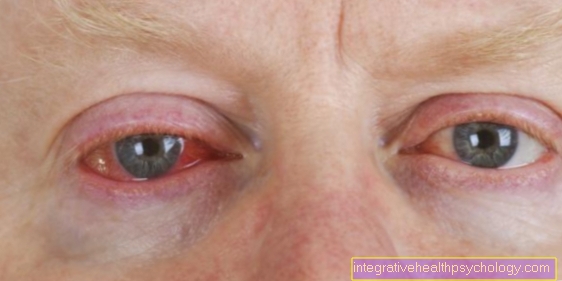
Endocrine orbitopathy
Endocrine orbitopathy is a condition that affects the eyes and their sockets (called the orbit). The majority of patients with endocrine orbitopathy develop this symptom as part of a thyroid dysfunction. The affected patient's eyes protrude from their sockets and the upper eyelids appear pulled up, making the eyes appear unnaturally large and wide open. The fact that it is still not possible for doctors to treat endocrine orbitopathy causally is due not least to the fact that the exact causes of the disease have not yet been fully researched.
Read more on this topic at: Endocrine orbitopathy
Sjörgen Syndrome
Sjögren's syndrome is an autoimmune disease in which the body's immune system v. a. directed against salivary and lacrimal glands. Sjögren's syndrome brings with it symptoms such as dry eyes, dry mucous membranes in the mouth, nose and throat as well as joint problems. Treatment has been difficult to this day due to the unexplained causality.
Read more on this topic at: Sjörgen Syndrome
Xanthelasma
Xanthelasma are yellowish plaques caused by lipid deposits in the upper and lower eyelids. They are harmless, in no case contagious and also not hereditary, although they can occur in families. In older people this often happens without a cause, in younger people underlying diseases must be ruled out. One recognizes the xanthelasma as yellowish cushions. If desired, cut out the affected areas of the skin.
Read more on this topic at: Xanthelasma
Rheumatic eye diseases
In rheumatic diseases, in principle all structures of the eye can be affected by inflammatory processes. One speaks of uveitis. Affected patients typically complain of burning eyes, a foreign body sensation, a reddened eye, stabbing pain and increased sensitivity to glare. Macular edema (swelling of the macula, the point of sharpest vision) or cataracts (cataracts, clouding of the lens of the eye) can occur as a complication of rheumatic disease.
In acute cases, rheumatoid diseases of the eye are treated with eye drops containing cortisone and, if necessary, with mydriatics. This is a pupil-widening medicine that is supposed to prevent the iris and iris from sticking together.
If the inflammation is frequent, there is a risk that the cortisone will cloud the lens of the eye. Therefore, in this case, so-called immunomodulatory therapy with methotrexate or cyclosporine A should be started, which suppresses the immune reaction.
Sjogren's syndrome is a special form of rheumatoid disease. It is an autoimmune disease that belongs to the type of collagenosis. In this disease, there is a change in the tear and salivary glands, which is why dry eyes and a dry mouth occur. Women after the menopause are particularly affected. The disease is treated with artificial tears and saliva. Eye drops containing cortisone or cyclosporine A can also be used.
Tumors and abnormalities in and around the eye

Choroidal melanoma
The choroidal melanoma is the most common malignant tumor inside the eye in adults. It is caused by a degeneration of the pigment-forming cells that are important for the color of the eyes. As a result, these tumors are often dark in color. The choroidal melanoma often metastasizes. In most cases, the choroidal melanoma does not cause any symptoms at first, which is why it remains undetected for a long time. The treatment of choroidal melanoma depends on the size and can be carried out using radiation, laser or surgery.
Read more on this topic at: Choroidal melanoma
Eyelid tumor
Lid tumors are growths of the eyelids. These can be both good and bad. The causes of the eyelid tumor include a wide variety of triggers. A high level of solar radiation (UV radiation) can promote the development of eyelid tumors. In addition, high exposure to X-rays has a negative effect. An eyelid tumor doesn't always have to be bothersome. Depending on the location, the tumors can also leave the patient completely unaffected. The treatment options for eyelid tumors depend on what type of tumor it is, how far it has progressed, where it is located and, consequently, what functional restrictions it brings with it.
Read more on this topic at:
- Eyelid tumor
- Conjunctival tumor
Retinoblastoma
A retinoblastoma is a tumor of the retina. This tumor is genetic, that is, hereditary. It usually occurs in childhood and is malignant. Retinoblastoma is a congenital tumor or it develops in early childhood. The affected children are actually symptom-free, i.e. they do not express any pain. Occasionally, children with retinoblastoma may squint. As already explained above, the retinoblastoma tumor is already well advanced at the time of diagnosis and is therefore relatively large. In these cases the eye must be removed. Smaller tumors can be treated with chemotherapy or radiation therapy.
Read more on this topic at: Retinoblastoma
Lacrimal gland tumors
Like all other organs, the lacrimal gland has both malignant and benign tumors. They differ in their growth pattern and their ability to scatter. The most common tumor of the lacrimal gland is that benign adenoma. Malignant tumors the lacrimal gland are rare. The therapy depends on the respective tumor.
Read more on this topic at: Lacrimal gland tumor
Birthmark in the eye
Colloquially, a well-defined, benign malformation of the skin or mucous membrane is called a birthmark or, in some cases, a pigment or mole spot. First of all, it is not a disease, but simply a benign abnormality. In rare cases, however, this can degenerate into melanoma. Usually it is only a cosmetic problem. It can be removed by surgery or laser coagulation.
Read more on this topic at: Birthmark in the eye
Cyst in the eye
A cyst describes a fluid-filled cavity that can appear anywhere on the body. A cyst in the eye describes a cavity in or around the eye that can be filled with sebum, pus, blood or tissue, for example.
Cysts in the eye can be congenital or acquired. Acquired means that the cysts were caused by infection, inflammation, or injury in the eye.
Typical locations for cysts in the eye are in the area of the conjunctiva, the iris (rainbow skin) and the lacrimal gland.
Since cysts are benign, they do not need to be removed until they cause symptoms such as pain or deterioration in vision.
In addition, there are more frequent cysts in the area of the eye, which are caused by an accumulation of sebum that has become encapsulated or through encapsulated sweat glands. They are known as sebum or sweat retention cysts.
Eye disorders due to nerve damage

Horner Syndrome
Horner's syndrome manifests itself through three defined signs of disease: pupil constriction, drooping of the upper eyelid and sinking of the eye into the eye socket. Horner syndrome is not a disease in its own right, just a symptom (sign) of a disease. Certain nerves are often damaged. A treatment of the Symptom Horner syndrome does not exist. However, treating the causes can reduce the signs of Horner's triad.
Read more on this topic at: Horner Syndrome
Optic atrophy
Optic atrophy is the loss of nerve cells in the optic nerve. The nerve cells either decrease in size or in number. Both are possible.
Atrophy can have various causes. The symptoms range from unnoticed, small central ones to large-scale and thus restrictive visual field defects in everyday life. The ophthalmoscopy by the ophthalmologist is particularly indicative in making the diagnosis. The treatment of optic atrophy is more difficult because the cause has to be treated.
Read more on this topic at: Optic atrophy
Vision disorders
Farsightedness
In farsightedness (hyperopia) there is an imbalance between the refractive power and the length of the eyeball. Far-sighted people see well in the distance, but objects appear blurry up close. The eyeball is too short in relation to the refractive power or the refractive power is too weak in relation to the eyeball. The cause can be axis hyperopia or refractive hyperopia. There are now several therapy options for correcting farsightedness. The simplest solution is glasses with convex lenses (also plus lenses or converging lenses) that support the refractive power of the eye.
Read more on this topic at: Farsightedness
Red Green weakness
The genetic red-green weakness is the most common color ametropia and is often incorrectly referred to as color blindness. It is always innate. Those affected perceive certain red and green tones only as shades of gray, which means that they are difficult or even unable to distinguish between these two colors. To date, there is no known therapy for red-green poor eyesight and since the disease is inherited, there are no options for prophylaxis.
Read more on this topic at: Red Green weakness
Squint
Squint is the deviation of an eye from the direction in which it should look naturally. There are various possible causes. Most of the time, however, no trigger can be identified. Symptoms include fatigue, headaches, and double vision. It can be treated with an operation. It is recommended to have the operation performed no later than 6 months from the time of occurrence.
Read more on this topic at: Squint
myopia
Nearsightedness (myopia) is a type of ametropia in which the relationship between the refractive power and the length of the eyeball is incorrect. Strictly speaking, the eyeball is too long (axial myopia) or the refractive power is too strong (refractive myopia). The short-sighted person can see objects in the vicinity well, but objects further away are only perceived as blurred or blurred. Myopia can usually be compensated for with the help of glasses.
Read more on this topic at: myopia
Night blindness
Night blindness is a disturbed ability of the eyes to adapt to darkness. For those affected, only outlines can be seen. It can also be acquired due to a vitamin A deficiency. At the ophthalmologist's office, night blindness is measured and recognized using devices. There is no therapy.
Read more on this topic at: Night blindness
Astigmatism / astigmatism
If you can see blurred both in the distance and in the vicinity, the cause may be a so-called astigmatism. The eye can no longer focus the incident light on an exact point on the retina and thus focus, but rather see points as blurred lines. A defective vision of the eye is called astigmatism. A distinction is made between regular and irregular astigmatism. The symptoms of astigmatism depend on the severity of the curvature of the cornea. In addition to correcting astigmatism with glasses or contact lenses, surgical intervention is a possible therapy.
Read more on this topic at: Astuteness and astigmatism
Color blindness
In the case of total color blindness, no colors, only contrasts (so light or dark) are perceived. A distinction is made between congenital and acquired color blindness. The main symptoms are: the inability to perceive colors; decreased visual acuity; quick, twitching eye movements; increased sensitivity to glare. There is currently no way to cure color blindness.
Read more on this topic at: Color blindness
Misalignments of the eye
Entropion
Entropion is a misalignment of the eyelid, more precisely an inward turning of the eyelid, so that the eyelashes drag on the cornea (trichiasis). This disease occurs mainly in older age (Entropion senile), but can also occur in babies. The permanent grinding of the eyelashes on the conjunctiva leads to reddening of the patient's eye and a feeling of foreign bodies.
Read more on this topic at: Entropion
Ectropion
Ectropion is a misalignment of the eyelid. But not inwards here (Entropion), but to the outside (Ectropion). In addition, the lower eyelid is almost always affected with ectropion. The lid is rolled outwards and often the inside shows up, as you can only see if you pull the lower lid down with your thumb. Ectropion - like entropion - is a disease of old age. Surgery is usually the method of choice. An attempt is made to surgically tighten the eyelid and reattach it to the eyeball, e.g. by shortening the lower eyelid and subsequent displacement.
Read more on this topic at: Ectropion
Psychosomatic eye diseases
It is not uncommon for people who are exposed to severe stress to report impaired vision. They often report that they see colors poorly, have problems reading, have dry eyes or cannot perceive part of their visual field (visual field loss).
There is also a disease called retinopathia centralis serosa, which occurs mainly in men under 50 years of age who are exposed to severe occupational stress. It is also known as a manager's disease. This is a detachment of the retina. There is an increased concentration of cortisol, a stress hormone, in the blood.
Genetic eye diseases
Common eye diseases that can be inherited are retinopathia pigmentosa, juvenile retinoschisis, congenital cataracts (congenital cataract) or congenital glaucoma (congenital glaucoma).
In retinopathia pigmentosa, the photoreceptors in the retina are destroyed. This leads to a gradual deterioration in vision.
Juvenile retinoschisis usually only affects men. However, women can still inherit the disease. One speaks of an X-linked recessive inheritance. The eyesight decreases continuously with the disease.
The glaucoma is an eye disease in which the intraocular pressure is increased. Only about 1% of those affected suffer from a hereditary form.
In cataracts, the lens becomes cloudy and, in the long term, impaired vision. Only some of those affected have congenital opacification of the lens, much more often it is caused by a natural aging process, diabetes mellitus or chronic eye inflammation.
Therapy of eye diseases
Vitamins for eye diseases
Studies have not shown that certain vitamins protect against eye diseases or improve existing eye diseases.
In general, vitamin A protects against free radicals and is mainly converted into carrots, spinach, broccoli, tomatoes and mangoes. The beta-carotene, which is partly contained in it, is also converted into vitamin A in the body.
Vitamins C and E are antioxidants and protect the body from cell damage. They are mainly found in citrus fruits, hazelnuts, whole grain products and vegetable oils.
Lutein is a component of the retina. It is therefore important for eyesight. This is mainly found in spinach and kale. It also contains zeaxanthin, which also has an antioxidant effect.
In general, the vitamins are said to strengthen eyesight and prevent macular degeneration or cataracts.