The function and piercing of the ear cartilage
Introduction - What is the ear cartilage?
There are many different types of tissue in the human body. One of these types of tissue is the cartilage and its sub-form, the elastic cartilage. This is located in the ear, among other things. The cartilage gives the outer ear its typical shape and ensures directed sound conduction into the ear. The cartilage also protects the ear canal. In children, the ear cartilage is still very soft, it only becomes firm in adolescence and takes on its characteristic shape. Inflammation or injuries can lead to a permanent change in shape, as the cartilage cannot renew (regenerate).

Function of the ear cartilage
Cartilage has a protective function throughout the body and occurs particularly in places with high mechanical stress. In the ear, too, the cartilage acts as a protection and shaping system for the sound-conducting system. The cartilage parts are arranged in such a way that a funnel results and the sound is optimally guided into the ear canal. This stabilization is particularly important inwards, as the ear cartilage merges directly with the ear canal cartilage. The ear cartilage gives the ear its characteristic shape and thus gives the human face a further individual factor.
The process of hearing begins with the characteristic shape of our ear. If you are interested in how things continue and ultimately how you can perceive tones and noises, we recommend our website: Listen
What can cause pain in the ear cartilage?
Ear cartilage pain can have different causes. The first group of causes are mechanical injuries. These are possible through external violence against the ear. These include both accidental knocks against the cartilage and piercing piercings. The physical abuse of children by pulling on their ears should also be considered in the case of cartilage injuries in children.The second cause of pain in the ear cartilage is inflammation and boils of the surrounding tissue structures. Boils, i.e. inflamed hair follicles that are filled with pus, can put pressure on the ear cartilage and cause pain. Mastoiditis, a serious complication of otitis media with bone involvement, also pushes the ear cartilage forward and causes pain. A one-sided protruding ear is noticeable in affected children. Another possible cause is neuralgia, i.e. nerve pain, after a herpes zoster infection in the facial area. Although there are no longer any foci of inflammation, the irritation of the nerves can persist for life.
If you have pain in the ear and can localize it more precisely, please read our pages for more detailed information and treatment options: Auricle pain or Ear entrance pain
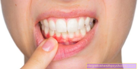
Ear cartilage inflammation
Like most types of tissue, the ear cartilage can become inflamed, but most inflammation originates in the cartilage membrane. A common cause is the pricking of piercings, as this creates an entry point for pathogens. Injuries are also possible. Mechanical irritation from cotton swabs can also lead to inflammation. Inflammation consists of several symptoms. The first symptom is Dolor, the pain that almost always occurs with inflammation. The second symptom is rubor, the redness, because inflammation leads to increased blood flow in the surrounding tissue. The overheating, called calor, is also due to the blood circulation. There may also be severe swelling in the area. The last main symptom is the Functio Laesa, i.e. the functional restriction. In the case of the ear cartilage, this is more due to the swelling, as this can narrow the ear canal. An inflammation of the ear cartilage should always be treated by a doctor, since once submerged ear cartilage does not renew (regenerate) and permanent changes in shape can occur. The treatment is carried out with disinfectants and therapy with antibiotics and cortisone.
You might also be interested in: Inflammation of the ear
Ear cartilage torn
The ear cartilage is a very elastic cartilage. In the case of "fractures" or tears in the ear cartilage, however, it is a matter of sudden giving in to external force with pointed or sharp objects. Since the cartilage cannot renew (regenerate), such injuries can permanently change the shape of the ear.
You might also be interested in: Torn earlobe
Piercing on the ear cartilage
Piercings are common on the ear. The most common locations are on the helix, i.e. on the outer edge of the ear. Tragus piercings are also common. However, the classic ear hole in the earlobe does not count among the cartilage piercings, as there is no cartilage there. Piercings in the ear cartilage can lead to cartilage inflammation, as a gateway for pathogens is created. After the piercing, severe swelling is to be expected and many piercing studios recommend antibiotic ointments or tablets in the first few days to weeks after the piercing.
If you are interested in stretching earlobes in the sense of a "tunnel" with a ring, we recommend you visit our website for more information: Ear lobe - piercing and tunnel
Piercing on the tragus
The tragus is the portion of cartilage closest to the face. It is a small protrusion of cartilage that comes from the face and lies over the ear canal, thus protecting it. The risk of cartilage inflammation is greater with tragus piercing than with other locations on the ear. The pain when stabbing should also be greater there.
Risks of piercing
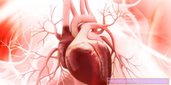
With piercings in the ear cartilage complications occur more often than with the classic earlobe piercing, since the cartilage skin is more sensitive than the soft tissue in the earlobe. The most common complication is inflammation. When piercing, an open wound is stabbed into the tissue and pathogens can get into the wound. This can happen during the piercing as well as afterwards. Cartilage inflammation should always be taken seriously, as otherwise it can spread to the middle and inner ear and, in the worst case, even to the brain. If it spreads via the blood, heart infections can also be triggered. Other diseases can also be transmitted when piercing. In the extremely rare case of non-sterile needles, infectious agents such as the HIV virus or hepatitis come. Depending on the location of the piercing, nerves can also be injured and permanent sensory disorders of the skin occur. When the piercing is inserted, an allergic reaction can also occur. This usually only happens when changing from surgical piercing to costume jewelry. Scarring with growths in the area of the puncture is also possible. In some people the piercing has to be removed because of the complications. After healing after twelve weeks, the risk of complications is significantly lower, but the risk of getting stuck and tearing the piercing remains permanently. This can lead to re-inflammation and the same complications as at the beginning.
Anatomy of the ear
The anatomy, i.e. the structure, of the ear is divided into a microscopic part and a part that is visible to the eye (macroscopic part). The microscopic part shows that the ear cartilage belongs to the elastic cartilage tissue. The elastic cartilage is a very cell-rich cartilage from only one Knoperle cell, in which hardly any groupings can be recognized. It also contains many elastic fibers that radiate into the cartilage skin. These elastic properties ensure great stability and hardly any vulnerability when bending (bendable) or pressing the ear. Otherwise our ear would be damaged on one side while sleeping, as the weight of the whole head presses on a small amount of tissue.
The macroscopic structure, i.e. the part that can be seen with the eye, consists of the outer ear and the outer ear canal (meatus acousticus externus). On the very outside, directed towards the back of the head, the helix lies in a large arc. Towards the face, the ear is bordered by the tragus. The second protruding arch, which is almost parallel to the helix, is called the antihelix and the lower end of this arch is called the antitragus. Other components are called Cavum Conchae, Crus helicis, Scapha and there are many more. The exact structure and shape of the individual parts varies greatly from person to person and also changes over the course of life.
For more information on the macroscopic structure of the ear, see: Outer ear, auricle and Earlobe
However, the ear is not only made up of the outer ear that is visible to us, but also of the middle and inner ear, which enables us to perceive noises and tones. For more information, we recommend our main page: The human ear


.jpg)