fetus
definition
Fetus or fetus translates as "descendant". A fetus is an unborn child in the womb. After fertilization, the developing child is called an embryo. When the internal organs are developed, the official name is fetus. The fetal period begins from the 9th week of pregnancy and ends with the birth. After birth, the fetus is called an infant.
Read more on the subject at: 1st trimester
development
Also read: Process of pregnancy & development of the embryo
1st - 4th week of pregnancy
In the first and second weeks of pregnancy, there is no pregnancy in the classic sense. The Egg cell is fertilized, but it has uterusin which it nests has not yet been reached. The Implantation in the uterus happens around the third week of pregnancy. The chromosomes on which the physical characteristics are stored are already determined at this point in time. So e.g. the eye or hair color, the gender is already determined genetically, but of course not yet recognizable.
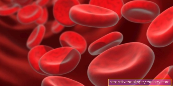
2 weeks after fertilization the egg cell accelerates cell division. After fertilization in the 3rd week of pregnancy the fertilized egg divides several times. In a short time, a larger cluster of cells is created while the body prepares the uterus for the egg cell to implant. After the cluster of cells has arrived in the uterus, it divides into 2 different areas. The one develops from one part placenta. It digs deep into the lining of the uterus. The embryo and later the fetus draw their nutrients from the placenta throughout pregnancy. The other part of the cell cluster develops into the embryo piece by piece. There is a vital connection between the developing child and the placenta: the umbilical cord. Parts of the placenta make up the pregnancy hormone beta HCG. It signals to the body that pregnancy has occurred and that ovulation does not currently have to take place. Most will use this hormone Pregnancy tests carried out.
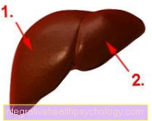
After implantation, the cluster of cells is initially divided into two different layers, from which the organs are later formed. One speaks of the outer and inner cotyledon. Endoderm and ectoderm. From the outer cotyledon In the course of the future fetus, the nervous system, the sensory epithelium, from which eyes, nose and ears are formed, skin and hair systems, sweat glands, mammary glands and tooth enamel develop. From the inner cotyledon develop the thyroid, liver and pancreas, almonds and thymus and the digestive tract. After creating these two cotyledons, a third one follows middle cotyledon (Endoderm). The so-called neural tube, from which most of the brain and nerves are formed, is formed by corresponding invagination. The middle cotyledon is divided into 2 larger symmetrical cell blocks. The following organs develop from them: the spine and vertebral bodies, bones and cartilage, the heart, muscles, connective tissue, blood and lymph vessels, the urogenital system and most internal organs.
5th week of pregnancy
The embryo is now about 2 mm in size and has already implanted itself in the uterus. From now on, growth and organ development begins. This stage of development will Embryonic phase called. From the 22nd day in the 5th week of pregnancy the embryo's heart begins to beat. Due to its small size, it must strike much faster than adults. On average, it hits 120-160 times a minute. The heartbeat is only detectable on the ultrasound from the 8th week. Furthermore, the head and torso are already put on in the 5th week.
6th week of pregnancy
The size of the embryo is about 4 mm. The neck and head can already be seen in the ultrasound. Eyes and ears are shadowy. The neck and thoracic vertebrae are clearly visible Ultrasound in pregnancy to recognize. The child's chest is formed from them in the further course.
7th week of pregnancy
The embryo is now about 5 mm in size.It will grow in size quickly in the following weeks. At this point you can clearly see your eyes and nose. The mouth can also be clearly seen as well as the brain structure. Hands and feet do not look fully developed and are not yet proportional to the rest of the body. Vessels shimmer through the thin skin (cannot be seen in the ultrasound) and the muscles that are applied develop rapidly. At this point the embryo begins to move.
8th week of pregnancy
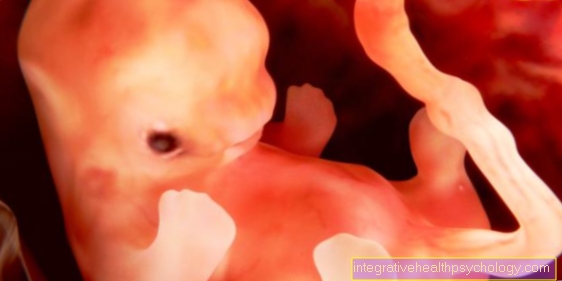
The embryo is in a hunched position. The head dominates almost the entire body, which is relatively small compared to the head. It now has a size of about 1.5 cm. The toes and fingers are now clearly visible and fully developed. The proportions gradually begin to equalize. Heartbeat and pulse are now clearly visible in the ultrasound. Many other organs are already completely set up and functional. Both kidneys are already in action and produce urine that is released into the amniotic fluid. The stomach begins to work and the upper body slowly straightens up. The joints of the upper and lower extremities begin to form, causing the embryo to stretch and bend the legs and arms. At this stage of pregnancy, however, movements are still uncontrolled and uncoordinated.
9-10 weeks of pregnancy
The embryo survived the first phase of development. The official name is now fetus. Despite the rapid development, the fetus is still in a vulnerable phase at this point in time. Abortions or miscarriages and recognizable malformations can occur. Numerous complications can arise. During the 9-10 week the internal organs are fully developed. The brain is completely covered by the skull bone. With the development of the brain and nerve tracts, the fetus can perceive external sensory impressions. So it can perceive vibrations, cold, warmth and pain. Furthermore, the fetus is practically in constant motion and kicks its arms and legs.
11th week of pregnancy
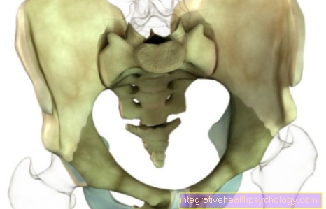
The size of the fetus is now about 4-5 cm. In addition to the complete organ arrangement and training, the external sexual characteristics (testicles and penis in boys, the vagina in girls) have now also developed. A point in time that is longed for by all parents-to-be, because from now on you can tell by an ultrasound scan whether it will be a boy or a girl. However, this statement is not yet entirely certain at this point in time. The gender can be seen more clearly around the 20th week of pregnancy. Another development stage in the 11th week of pregnancy is the enclosure of the rib cage around the internal organs to protect them. At this point in time, all organs are in place and either still in development or development has already been completed. Tooth roots are laid out in the upper and lower jaw as well as hair roots.
12th week of pregnancy
This ends the first trimester of pregnancy (1st trimester) and thus the "dangerous phase" for the fetus. Statistically, the fetus is now less threatened by serious events such as an abortion or malformations. Miscarriages can still occur, but since the majority of miscarriages occur due to chromosome damage, which particularly affects the first developmental steps, the risk is significantly reduced. In most cases, the parents-to-be also inform relatives and friends at this point.
It is still important to be appropriate Check-ups during pregnancy to have an ultrasound carried out and to continue to secure the pregnancy in order to recognize dangerous processes at an early stage.
The fetus is now about 5 cm long, is moving vigorously and is active. He uses arms and legs for movement and moves his head. The size of the fetus is measured by an ultrasound examination using a line connecting the skull and the rump (buttocks of the fetus) (skull-rump length). The weight of the fetus is also calculated by the ultrasound and is usually around 16 grams in the 12th week of pregnancy. The head is still disproportionately large, but it is slowly beginning to adapt to the body. The eyes also move to the front of the head. At this point in pregnancy, the fetus is still completely blind. Throughout pregnancy, nutrients and oxygen are supplied through the umbilical cord, which is connected to the mother via the placenta. The placenta is also a filtering organ, but some toxins are also let through. It is accordingly It is very important that the expectant mother does not consume alcohol or smoke. Even when taking various medications, a doctor should be consulted beforehand and asked about the harmlessness of taking them.
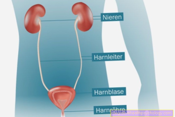
The amniotic fluid in the amniotic sac acts as a protective buffer and protects the fetus from shocks and movements, but it also serves as a "waste water container", as the urine of the fetus is released into the amniotic fluid. At the time of the 12th week of pregnancy, the female external sexual organs also become visible. Here, too, it is not yet possible to make a definitely reliable diagnosis as to whether it will be a boy or a girl. The gynecological check-up during pregnancy takes place every 4 weeks up to the 32nd week of pregnancy. Thereafter, the interval is reduced to every 2 weeks. Thus, within a pregnancy 10-12 examination appointments scheduled. In addition to the ultrasound control of the fetus, urine and blood pressure checks of the mother, as well as weight checks and general physical examinations of the mother are carried out. In addition, the heart rate and the position of the child are determined in advanced pregnancy. Overall are during pregnancy three major ultrasounds planned. The first takes place between the 9th and 12th week of pregnancy. It represents the basic examination. The second examination, which can also be carried out as an extended ultrasound examination, should be carried out between the 19th and 22nd week of pregnancy. The last one between the 29th and 32nd week of pregnancy. If you have problems or complaints, you can of course do an ultrasound at any time.