Heel bone
anatomy
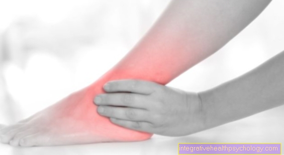
The heel bone (lat. Calcaneus) is the largest and dominant foot bone and has a slightly cuboid shape. As part of the rear foot, part of the heel bone stands directly on the floor and serves for stability. The calcaneus is divided into different parts, which fulfill different functions and tasks.
More about the heel can be found here: Achilles heel

- Toe phalanx -
Phalanx distalis - Middle toe -
Phalanx media - Phalanx -
Phal. proximalis
(1st - 3rd toe bones -
Phalanges) - Metatarsal bones -
Os metatarsi - Inner sphenoid bone -
Medial cuneiform bone - Middle sphenoid bone -
Os cuneiform intermedium - External sphenoid bone -
Os cuneiform laterale - Cuboid bone - Os cuboideum
- Scaphoid bone - Navicular bone
- Ankle bone - Talus
- Ankle roll - Trochlea tali
- Heel bone - Calcaneus
- Protrusion on the 5th metatarsal -
Tuberositas ossis metatarsalis quinti (V)
You can find an overview of all Dr-Gumpert images at: medical illustrations
The posterior prominent portion of the calcaneus is called the Calcaneal tuberosity and is visible and palpable as the heel on the foot. This is where the Achilles tendon, of the Twin calf muscle (Gastrocnemius muscle) and the Clod muscle (Soleus muscle) on. On its underside, a stabilizing band runs between the heel bone and the cuboid bone (Calcaneocuboid ligament). In addition, there are two cusps on the underside, the processus lateralis tuberis calcanei and the Processus medialis tuberis calcanei. These serve as the origin for the Abductor hallucis muscle, the Flexor digitorum brevis muscle and the Abductor digiti muscle minimi.
Also the tendon plate in the area of the sole of the foot, Plantar aponeurosis, has its origin in the calcaneal tuberosity. To the front, the calcaneus forms with the cuboid bone (Os cuboideum) an articulated connection. Both on the inside of the foot and on the outside of the heel bone are protruding bones that protect and guide muscles. The is on the inside of the foot Sulcus tendinis musculi flexoris hallucis longuswhich contains the long flexor muscle of the big toe and prevents the heel bone from buckling inwards through its action. This is covered by a protruding bone, the Sustentaculum tali.
On the outside of the foot is the Sulcus tendinis musculi peronei longi. This muscle is used to tension the Transverse vault. In addition, various nerves and blood vessels run in these boxes.
On the top of the calcaneus there are three articular surfaces, the Facies articularis talaris anterior, Facies articularis talaris media and the Facies articularis talaris posterior. The runs between the last two joint surfaces Calcaneal sulcus, which together with the Talar sulcus of the talus as Canalis tarsi designated tunnel forms. The anterior (anterior) and middle (medial) articular surfaces are parts of the anterior ankle.
The rear (posterior) articular surface is part of the posterior ankle. The entire heel bone and especially the prominent rear portion is the decisive pressure point for standing and walking upright.
Appointment with ?

I would be happy to advise you!
Who am I?
My name is dr. Nicolas Gumpert. I am a specialist in orthopedics and the founder of .
Various television programs and print media report regularly about my work. On HR television you can see me every 6 weeks live on "Hallo Hessen".
But now enough is indicated ;-)
Athletes (joggers, soccer players, etc.) are particularly often affected by diseases of the foot. In some cases, the cause of the foot discomfort cannot be identified at first.
Therefore, the treatment of the foot (e.g. Achilles tendonitis, heel spurs, etc.) requires a lot of experience.
I focus on a wide variety of foot diseases.
The aim of every treatment is treatment without surgery with a complete recovery of performance.
Which therapy achieves the best results in the long term can only be determined after looking at all of the information (Examination, X-ray, ultrasound, MRI, etc.) be assessed.
You can find me in:
- Lumedis - your orthopedic surgeon
Kaiserstrasse 14
60311 Frankfurt am Main
Directly to the online appointment arrangement
Unfortunately, it is currently only possible to make an appointment with private health insurers. I hope for your understanding!
Further information about myself can be found at Dr. Nicolas Gumpert
Injuries and pain in the heel
The most common injuries to the calcaneus are Fractionscaused by falls from a great height or traffic accidents. The patients suffer from very severe pain and can no longer stand or walk as a result. Of the Fracture of the calcaneus is divided into different degrees of severity. Fractures with joint involvement (intra-articular) heal significantly worse and require surgical treatment. On the other hand, fractures lying outside the joint (extra-articular) can usually conservative can be treated with immobilization and pain medication.
Read more on the topic:
- Heel bone pain
- Heel pain
- Haglund heel
- Heel spur


