Vascular supply diaphragm
General
The diaphragm is the most important respiratory muscle and separates the chest from the abdomen.
Figure diaphragm
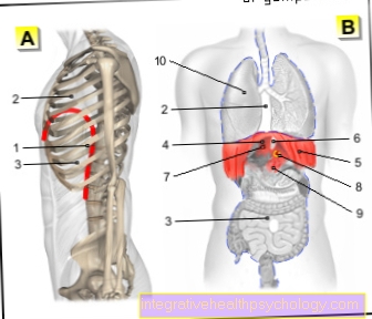
- Diaphragm (red) -
Diaphragm - Chest cavity -
Cavitas thoracis - Abdominal cavity -
Cavitas abdominis - Tendon center of
Diaphragm -
Centrum tendineum - Rib part of the diaphragm -
Pars costalis diaphragmatis - Esophageal slit -
Aortic hiatus - Vena cava hole -
Foramen venae cavae - Aorta in the aortic slit -
Aorta in the aortic hiatus - Loin part of the diaphragm -
Pars lumbalis diaphragmatis - Lungs - Pulmo
The diaphragm separates
the thoracic and abdominal cavities
You can find an overview of all Dr-Gumpert images at: medical illustrations
Arterial supply
The arterial supply (Vascular supply to the diaphragm) is complex and takes place via four different branchesthat branch out heavily. First of all, it is the upper diaphragmatic arteries (Arteriae phrenicae superiores), the Diaphragmatic sac artery (Pericardiacophrenic artery) and the Diaphragmatic artery (Musculophrenic artery), all from the Thoracic artery (Thoracic aorta) arise from. The further arterial supply takes place via the lower ones Diaphragmatic arteries (Inferior phrenic arteries) from the Abdominal artery (Abdominal aorta) originates (vascular supply diaphragm).
Venous outflow
The venous outflow takes place via veins of the same name into the superior vena cava (Superior vena cava). Only the lower diaphragmatic veins (Inferior phrenic veins) opens into the inferior vena cava (Inferior vena cava) (Vascular supply diaphragm).