Joints of man
Synonyms
Joint head, joint socket, joint mobility,
Medical: Articulatio
English: joint
Number of joints
The number of joints in a person depends on whether you add up only real joints or all articulated connections in the body.
There are around 100 real joints in the human body, i.e. joints that consist of two joint partners, are separated from one another by a joint space lined with cartilage and have a joint capsule.
If you add any articulated connections, i.e. all structures connected by ligaments, tendons or cartilage that allow movement, you get a number of around 360 articulated connections.
For many people this is an astonishingly high number, since the most well-known joints are only dated to a number of six joints per side of the body, i.e. twelve joints (shoulder-, Elbow-, hand-, Hip-, knee- and Ankle).
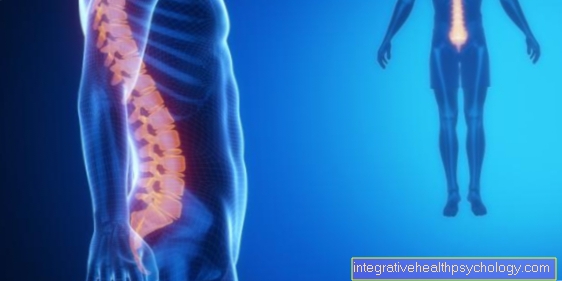
The far more numerous joints skull, Spine, the Hands and Feet are often not so conscious of the human being. The joints on the trunk in particular are not consciously moved and are not as clearly visible as the large joints on the extremities.
Nevertheless, they are essential for the mobility and flexibility of the human body.
The individual joints of the person
Inner collarbone joint
The inner collarbone joint (Art. Sternoclavicularis) consists of the articular surfaces of the
- Collarbone (Clavicle) and des
- upper sternum part (Manubrium sterni).
They are both slightly saddle-shaped and do not fit perfectly. This is balanced out through a discus. The joint is secured and mobility is restricted by tight straps. These are that
- anterior and posterior collarbone sternum ligament (Ligg sternoclaviculare anterius and posterius)
- the ligament between the two clavicles (lig. interclavicular) and
- the rib clavicle ligament (lig costoclavicular).
The inner collarbone joint is the only bony connection between the shoulder girdle and the chest. The two main movements are forward and backward, and shoulder raising and lowering. In addition, the collarbone can be rotated around its longitudinal axis.
Figure joint shapes
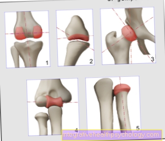
- Wheel angle joint
= Swivel hinge joint
(e.g. knee joint) - Saddle joint
(e.g. thumb saddle joint) - Ball joint
(e.g. shoulder joint,
Hip joint) - Hinge joint
(e.g. elbow joint) - Wheel joint
= Pivot joint
(e.g. spoke-ulnar joints) - Egg joint (not shown)
similar to ball joint,
only biaxial
(e.g. proximal wrist)
Uniaxial Joints -
Hinge joint and wheel joint
Biaxial joints -
Wheel angle joint, saddle joint
and egg joint
Triaxial joint - Ball joint
You can find an overview of all Dr-Gumpert images at: medical illustrations
External clavicle joint
The outer collarbone joint (Art. acromioclavicularis) is also known as the shoulder joint. It is the connection of the shoulder roof (Acromion) with the collarbone (Clavicle) and a flat joint that is supported by three tight ligaments,
- the shoulder collarbone strap (Acromioclavicular ligament)
- the raven-beak collarbone ligament (Coracoclavicular ligament) and
- the raven beak extension shoulder roof band (Coracoacromial ligament).
There are shifting movements forwards, backwards, upwards, downwards and a rotation of the collarbone around its own axis. Also read ankle joint dislocation
Shoulder joint
The shoulder joint (Art. humeri) is the most flexible and vulnerable joint in the body. It is made up of:
- the head of the humerus (Caput humeri) and
- the socket of the shoulder blade (Glenoid Cavitas).
The joint surface is three to four times smaller than the joint head, which allows great mobility but also low stability.
The shoulder roof (Fornix humeri) serves as an additional safeguard for the head in the pan. This roof consists of:
- the shoulder roof (Acromion)
- the raven beak process (Proc. coracoid) and
- the raven-billed roof tape (Lig. coracoacromialis).
The shoulder joint capsule is wide and very thin at the back. On the front is the capsule with ribbons (Glenohumeral ligament) reinforced. If the arm hangs down, a lower bulge forms (Axillary recess), which allows great mobility. The joint capsule is with neighboring bursa (Bursa subtendinea musculi subsacapularis and the subcoracoid bursa) and the tendon sheath of the long biceps tendon runs inside the capsule. Three degrees of freedom with six main movements are possible in the shoulder joint:
- the spreading (Abduction) and
- Bring up (Adduction),
- the bowing (Flexion) and
- Stretch (Extension) and the
- Internal rotation and
- Outward rotation.
Elbow joint
The elbow joint (Art. cubiti) is a composite joint made up of three partial joints:
- the upper arm joint (Art. humeroulnaris),
- the elbow joint close to the body (proximal radioulnar joint) and
- the upper arm spoke joint (Art. humeroradialis).
The upper arm joint is a hinge joint with one degree of freedom and two directions of movement, flexion and extension. The structure of the upper arm spoke joint is a ball joint, whereby only two degrees of freedom are possible due to the band-like structures. In addition to the flexion and extension, which takes place together with the upper arm spoke joint, the joint also enables inward and outward rotation (Per- and Supination) of the forearm.
The ulnar spoke joint near the body is a flat joint where the ulna and radius move. Three ligaments are crucial in the elbow joint.
- the inner sideband (Lig collaterale ulnare) and
- the outer sideband (Collateral radial ligament) stabilize the joint and
- the ring band (Annular radii ligament), which runs in a ring around the head of the spoke and secures it in the joint.
wrist
The wrist is made up of two joints.
- on the one hand, close-fitting wrist (Art. Radiocarpea) and
- the distant wrist (Art. Metacapea).
The wrist joint close to the body is an egg joint with two degrees of freedom, the socket of which is formed from the spoke, a disc that evenly distributes the pressure forces and a stylus extension of the ulna. The head is formed from the scaphoid bone, the lunar bone and the triangular bone of the carpal bones.
The wrist distant from the body is made up of the carpal bones mentioned above and the remaining carpal bones, hook bone, head bone, small polygonal bone. The joint gap is S-shaped so that both rows of carpal roots are interlocked. One speaks here of a toothed hinge joint. Both joints form a functional unit when moving. The movements of the wrist are flexion and extension and lateral splay. There are tight ligament connections between the carpal bones (Amphiarthroses).

Saddle thumb joint
The thumb saddle joint (Art.carpometacarpalis polis) consists:
- the large polygon and
- the first metacarpal bone.
It is a saddle joint with three degrees of freedom and thus six movements are possible, the flexion, extension, spreading and bringing closer and additionally the opposition and reduction movement to the little finger.
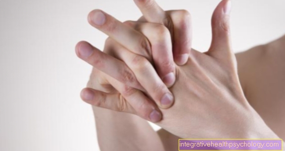
Finger joints
The finger joints (Articulationes digitorum) are in:
- Basal joints
- Central joints and
- Subdivided end joints.
The basic joints (Articulationes metacarpophalangeae) are located between the heads of the metacarpal bones and the bases of the phalanges close to the body. Both joint surfaces are pan-like and they are ball joints with two degrees of freedom. Bending, stretching, spreading and bringing up are possible. The finger joints near and far from the body (Articulationes interphalangeales proximalis and distalis) are hinge joints with one degree of freedom and two movements, flexion and extension. All carpal bones are connected by numerous ligaments. In addition, ligaments pull the forearm and metacarpal bones. The ligaments strengthen the joint capsules in the upper areas. They are divided into four groups according to their location and arrangement: the ligaments between the forearm and carpal bones, the ligaments between the carpal bones, the ligaments between the carpal and metacarpal bones and the ligaments between the bases of the metacarpal bones.
Sacrum-iliac joints
The sacrum-iliac joints (Articulationes sacroilacae) is made from the two ear-shaped articular surfaces of the iliac bone (Os ilium) and sacrum (Sacrum) educated. The cartilage surfaces are mountainous and therefore well wedged into one another, so that only small movements, the tilting forward (Nudation) and the erection (Counter-nudation) of the sacrum are possible. The ligaments that secure the tight joint capsule are in front:
- the anterior sacrum-iliac ligament (Lig. Sacroiliacae ventralia) and back
- the posterior sacrum-iliac ligament (Lig. Sacroiliacae dorsalia) and the inter-bony sacrum-iliac ligament (Lig. Sacroiliacae interosseus).
- In addition, the joint is supported by the iliac ligament (Lig. Iliolumbar) between the iliac crest and the last lumbar vertebrae,
- the sacrum seat hump ligament (Sacrotuberous ligament) from the sacrum to the seat hump and the
- Sacrum lace ligament (Sacrospinal ligament) from the sacrum to the tip of the ischium.
hip joint
The hip joint (Art.coxae) consists:
- the acetabulum (Acetabulum) and the
- Femoral head (Caput ossis femoris).
The hip joint is a nut joint. The joint surface of the socket is crescent-shaped (Facies lunatum) and encloses a pit filled with fatty tissue (Acetabular fossa). The articular surface is bordered by a bony edge (Limbus acetabuli), on which a cartilaginous joint lip sits. This cut (Acetabular notch), which is from a tape (Lig. Transversum acetabuli) is spanned. All of these structures ensure that the joint surface surrounds the joint head like a nut and includes freedom of movement. The joint capsule is relatively wide and envelops the head and most of the femoral neck.
It arises at the bony edge of the joint socket and runs to the intercuspid line (Linea intertrochanterica or Crista intertrochanterica
- the iliac-thigh ligament (Lig. Iliofemoral) from the posterior edge of the socket to the trochanteric fossa (Trochanteric fossa),
- the ischio-femoral ligament (lig. ischiofemorale) from the posterior edge of the socket to the trochanteric fossa and
- the pubic-thigh ligament (lig. pubofemoral) from the upper pubic branch and radiates into the features of the iliac-thigh ligament.
These three bands run helically and secure the head in the pan. The femoral head ligament runs inside the joint capsule and pulls from the femoral head recess (Fovea capitis) to the socket of the joint socket (Acetabular fossa). It has no stabilizing function, but serves as a vascular ligament for nourishing the femoral head. With three degrees of freedom, the hip joint has six directions of movement: flexion, extension, approaching and spreading and turning in and out.
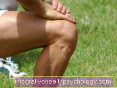
Knee joint
The knee joint (Art. Genus) is the largest joint in the human body. It is a compound joint and is made up of the bones
- Shin (Tibia)
- Thigh (Femur) and
- Kneecap (patella).
The shin and thigh together form the shin-thigh joint (Art. Tibiofemuralis), while the thigh and kneecap together form the kneecap-thigh joint (Art. Patellofemuralis) form. The two joints are encased in a joint capsule and are located in a joint cavity.
In the shin-thigh joint, the two spherical extensions of the thigh (Condyles) and the hollowed plateau of the tibia (Tibial plateau) the articular surfaces. In between are the two menisci to compensate for unevenness between the two joint partners and to absorb pressure.
Since there are two menisci, another distinction is made between two partial joints, the right and left meniscus tibial joint and the right and left meniscus thigh joint. There is a hump between the two articular surfaces of the tibial plateau (Eminentia intercondylaris) to which the cruciate ligaments and the two menisci attach. In the thigh kneecap joint, the kneecap and the thigh form the two joint partners. The base of the kneecap is round while at the bottom it tapers into a point. The articular surface covered with cartilage is traversed by a ridge so that it can slide between the two thigh processes like on a splint.
The joint capsule stretches from the tibia plateau to over the two thigh processes. The kneecap and the kneecap tendon are embedded in the anterior wall of the capsule. The joint capsule is connected to the neighboring bursa in many places, so that the capsule can unfold completely with all movements and the kneecap allows undisturbed gliding.
The ligament apparatus consists on the one hand of the two lateral ligaments. The inner ligament runs from the rear above the inner thigh process to the inside-front on the side of the tibial plateau. It lies directly on the capsule and is fused with it and the meniscus below. The outer collateral ligament runs from the front above the outer thigh process to the fibula head. It is not associated with the capsule.
The two side ligaments lock the knee joint in the extended position of the knee joint so that no shear stress is permitted. The two cruciate ligaments lie within the joint capsule, but are located between the two layers of the joint capsule.
The anterior cruciate ligament comes in front from the tibial plateau and pulls to the inner surface of the outer thigh process, while the posterior cruciate ligament at the back pulls from the tibial plateau to the inner surface of the inner thigh process. They enable contact between both joint partners in every joint position and prevent inward rotation when the knee is extended. Two degrees of freedom with four movements are possible in the knee joint
- Bend and
- Stretch and that
- Turning in and out.
Tibia-fibula joint
The shin-fibula connections are the proximal and the distal shin-fibula joint (Art. Tibiofibularis proximales et distales). They are plane joints that can only be shifted. The distal tibial-fibula joint also plays a crucial role in the movements of the upper ankle. It forms the so-called ankle fork and stabilizes the upper ankle joint. Both joints are held together by tight ligaments.
upper ankle
The upper ankle (Art.talocruralis) sometimes also called the ankle joint from the distal ends of the tibia and fibula as well as from the ankle roll (Trochlea tali) of the talus (Talus) educated. This joint is the place where power is transmitted from the foot to the lower leg. The joint capsule arises from the cartilage-bone boundary and is thin and flexible in the front area. It is reinforced at the front by connective tissue structures that fix the tendons of the lower leg muscles.
The capsule is reinforced by ribbons on the back and sides. The outer ligaments are the anterior and posterior ankle-fibula ligaments (Lig. Talofibular anterius et posterius) and the calcaneus-fibula ligament (Calcaneofibular ligament). The inner band is also called a triangular band (Deltoid ligament) and consists of four parts,
- the anterior and posterior tibial ankle parts (Pars tibiotalares anterius et posterius),
- the shin-scaphoid part (Pars tibionaviculare) and the
- Shin-calcaneus part (Pars tibiocalcanea).
The ankle joint is a joint with one degree of freedom and thus two directions of movement, the
- Diffraction and
- Elongation
lower ankle
The lower ankle (Art. Talotarsalis) is a compound joint. Here articulate the talus (Talus) with the calcaneus (Calcaneus) and the navicular bone (Navicular bone). A distinction is made between two completely separate partial joints, which are called so-called
- rear joint chamber (Art. Subtalaris) and
- anterior joint chamber (Art. Talocalcaneonaviculare)
The ankle bone and heel bone articulate in the posterior joint chamber, while the ankle bone articulates in the anterior joint chamber with a socket made of the calcaneus, scaphoid and the so-called acetabular ligament. The acetabular ligament is a crucial ligament structure that contributes to the formation of the longitudinal arch. The joint capsule is thin and wide and is formed on the one hand by the acetabular ligament and on the other hand by the strong interbone-talus-calcaneus ligament running in the joint (Lig. Talocalcaneum interosseum). This ligament connects the talus to the calcaneus and separates the joint into the two chambers. The ligament guides vessels that supply the talus.
Inside, outside and behind the anterior chamber of the lower ankle joint is defined by the inner, outer and posterior ankle-calcaneus ligament (Ligg. talocalcaneum mediele, laterale et posterius) stabilized. The joint capsule of the anterior chamber is attached to the rear by posterior ankle-navicular ligament (Talonavicular dorsal ligament).
Outside, a V-shaped band runs from the calcaneus to the navicular bone and to the cuboid bone (Lig. Bifurcatum). The lower ankle joint causes a possible twisting of the foot.
These are other joints of the foot
- Calcaneal-cuboid joint (Art. Calcaneocubuidea),
- the transverse tarsal joint or Chopart joint (Art. Tarsi-transversa),
- the sphenoid-scaphoid joint (Art. Cuneonavicularis),
- the joints between the sphenoid bones (Articulationes intercuneiformes),
- the joint between the outer sphenoid bone and the cuboid bone (Art. Cuneocuboidea)and
- the tarsal-metatarsal joints or also Lisfranc joints.
The Chopart joint are the joint lines of the navicular-calcaneus and the calcaneal-cuboid joint. With the help of this joint, the forefoot can be moved in flexion, extension and rotation in relation to the rear foot. All other joints are spurious due to tight ligament connections.
Joint chamber
The toe joints are transformed into the metatarsophalangeal joint (Art. Metatarsophalangeae) and in the middle and end joints (Art. Interphalangeae proximales et distales). The metatarsophalangeal joints consist of the cylindrical head of the metatarsal bones and the joint socket at the bases of the first toe bones and are enclosed by a wide joint capsule. The movements are like the movements of the metatarsophalangeal joints
- Diffraction,
- Stretching and that
- Approach and apart and the
- rotation
The basic joints are functionally enhanced by tight collateral ligaments (Ligg. collateralia) to hinge joints. On the sole of the foot, the joint capsule is secured by tight ligaments (Ligg. plantaria) reinforced. The middle and end joints are classic hinge joints that allow flexion and extension. The strongest band on the soles of the feet is the soles of the feet (Plantar ligament), which is important for tensioning the longitudinal arch.
Appointment with ?

I would be happy to advise you!
Who am I?
My name is dr. Nicolas Gumpert. I am a specialist in orthopedics and the founder of .
Various television programs and print media report regularly about my work. On HR television you can see me every 6 weeks live on "Hallo Hessen".
But now enough is indicated ;-)
In order to be able to treat successfully in orthopedics, a thorough examination, diagnosis and a medical history are required.
In our very economic world in particular, there is too little time to thoroughly grasp the complex diseases of orthopedics and thus initiate targeted treatment.
I don't want to join the ranks of "quick knife pullers".
The aim of any treatment is treatment without surgery.
Which therapy achieves the best results in the long term can only be determined after looking at all of the information (Examination, X-ray, ultrasound, MRI, etc.) be assessed.
You will find me:
- Lumedis - orthopedic surgeons
Kaiserstrasse 14
60311 Frankfurt am Main
You can make an appointment here.
Unfortunately, it is currently only possible to make an appointment with private health insurers. I hope for your understanding!
For more information about myself, see Lumedis - Orthopedists.


.jpg)