ureter
Synonyms
- Urinary tract
- Primal entrance
- kidney
- bladder
Medical: Ureter
English: ureter
anatomy
The ureter connects the renal pelvis (pelvis renalis), which collects urine from the kidney like a funnel, with the urinary bladder.
The ureter is a 30-35 cm long tube made of fine muscles and about 7 mm in diameter.
It runs behind the abdominal cavity (abdomen) on the inner back muscles down into the pelvis, where it reaches the urinary bladder from behind.
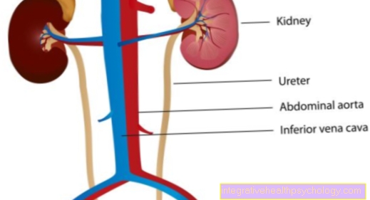
The right ureter is a little shorter because the right kidney is slightly lower due to the space-consuming expansion of the liver. The ureter joins the urinary bladder at an angle, which is beneficial for the closure of the ureter because it is compressed by the strong muscles of the urinary bladder so that, for example, urine cannot flow back into the ureter when lying down.
In addition to this narrow point at the end of the ureter, two more arise on the way to bladder. The transition from the renal pelvis to the ureter shows a narrowing, just as the clearing of the ureter is narrowed by the large blood vessels in the pelvis when the ureter enters the pelvis. These three constrictions can play an important role if there are stones in the ureter (Kidney stones), which can then get stuck (see below).
The ureter is in the pelvis in women cervix (Cervix uteri) and in men to the spermatic duct (ductus deferens) adjacent.
Figure ureter
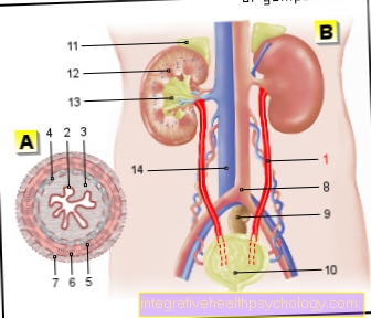
- Ureter - Ureter
- Transitional epithelium - Urothelium
- Shift layer of the
Mucous membrane - Lamina propria - Inner longitudinal layer -
Stratum longitudinal internum - Outer longitudinal layer -
Stratum longitudinal externum - Middle ring layer -
Circular stratum - Connective tissue covering with
Blood vessels - Tunica adventitia - Aortic fork - Aortic bifurcation
- Rectum - Rectum
- Urinary bladder - Vesica urinaria
- Adrenal gland -
Glandula suprarenalis - Right kidney - Ren dexter
- Renal pelvis - Pelvis renalis
- Lower vena cava - Inferior vena cava
You can find an overview of all Dr-Gumpert images at: medical illustrations
function
In addition to its function as a link between kidney and bladder, the ureter also has an important role in moving the Urine. When lying down, gravity counteracts the flow of urine.
The ureter can gradually tense its muscles so that the urine reaches the urinary bladder against the gradient like on a conveyor belt.
This post-and-post tensioning is called a peristaltic wave. It runs 1-4 times per minute via the ureter. The principle is similar to that of esophagus, which is also food for the headstand stomach promoted.
Diseases of the ureter
Ureteral stone, urinary stones, kidney stones in general
Men and women are of Kidney stones affected equally often.
With age, the risk of developing a stone in the kidney increases. Stress can adversely affect the occurrence of urinary stones / kidney stones.
The climate can also have an impact on the development of kidney stones. The more water is lost through sweating, the more concentrated the urine.
If the urine is too concentrated or certain substances are in abundance, possibly due to poor nutrition or certain congenital disorders in the breakdown of waste products in the body, the likelihood of urinary stones is greatly increased because these substances can no longer dissolve in the urine and fall out as crystals . Here the so-called PH value, so the acidity, the urine plays an important role. Depending on how much acid there is, some stones form more easily.
A inflammation in the urinary system or if the outflow of urine is disturbed, for example by congenital malformations, can also promote the formation of urinary stones.
Normally the body produces substances that inhibit stone formation. If there is too little of it, urinary stones can form more easily. You can distinguish between different stones based on their composition and origin.
On the one hand, urinary stones / kidney stones can arise in the renal pelvis (pelvis renalis) anchored to the wall. These are called chalices or fixed stones. They can loosen and be washed into the urinary drainage system in the ureter. On the other hand, uric acid and cystine stones arise freely in the urine, simply because the concentration of these substances is too high or because the pH value of the urine has changed. They can arise anywhere in the urinary system.
Most stones (70%) are made of calcium oxalate, if too much calcium or oxalate is present in the urine or there are not enough anti-stone substances.
Uric acid stones (10-15%) arise when purine accumulates. Purine is a breakdown product of, for example, DNA, which we ingest in large quantities when we eat meat. If the breakdown is disturbed, possibly due to a congenital defect or if the kidney is damaged or is overwhelmed by an excessive intake of meat and alcohol, these stones arise.
Calcium and magnesium phosphate stones (5-10%) are so-called Infection stoneswhich are formed when bacteria change the pH of the urine through their waste materials when they become infected.
Cystine stones are rare (1-2%) and are mainly composed of the protein component cystine. They mostly arise due to a hereditary enzyme deficiency.
Xanthine stones and other stones make up less than 0.5% of all kidney stones.
People with urinary stones will mainly become aware of the stones when they are in the ureter and cause pain due to the stretching of the ureter wall.
These pains are mostly colic-like (that is, they come and go in the form of waves) with a wave-like spread in the flanks, in the urinary bladder or in the Scrotum (Scrotum) in men or those Labia (Labia majora) in women.
In addition, if the urine is blocked, the urge to urinate cannot be resolved. If the urine congestion persists, it may cause inflammation or a Blood poisoning with urine substances that cannot be excreted (Urosepsis).
Ureter stones (ureter stones) can be found mainly through imaging procedures such as Ultrasonic or contrast agent examinations (intavenous urogram).
Ultrasound can detect stones that are larger than 2 mm. But the urine test can also provide an indication of the presence of blood or small urinary stone crystals.
Depending on the crystals discovered and the pH value, conclusions can also be drawn about the cause.
A blood test can also be informative if so-called urinary substances such as creatinine occur more frequently.
Since 70-80% of the stones come off spontaneously because they are propelled by the peristaltic wave of the ureter described above, one can usually use an anticonvulsant such as Buscopan® and treat pain relievers.
Uric acid stones, which sometimes arise due to the acidity, you give alkalizing drugs that neutralize the urine a little and thus dissolve the stones, e.g. Uralyt U (this is the salt of citric acid).
If the stones cannot be dealt with with medication, one can resort to so-called endourological measures, which are characterized by inserting a special catheter through the ureter past the stone and allowing the blocked urine to drain away. The stone is usually pushed back into the renal pelvis, where it can be smashed more easily (see below).
Stones can be smashed by a special technique from the outside by certain radio waves or electromagnetic waves without having to intervene directly in the body (extracorporeal shock wave lithotripsy). No general anesthesia is required and the small debris can easily be expelled through the ureter and bladder.
In the case of very stubborn or large stones, invasive access to the stone must also be created through the skin (percutaneous nephrolitholapaxy).
Since ureteral stones are particularly difficult to locate, they are usually treated endoscopically under anesthesia. That means you lead a hose equipped with a camera over the urethra (Urethra) that bladder (Vesica urinaria) into the ureter and can then precisely remove the stone with the help of the image.
You can prevent the formation of urinary stones if you adjust your diet accordingly, exercise a lot and drink plenty of fluids. You can also take magnesium and citrate to prevent the formation of stones. In the case of infection stones, L-methionine is often given a protein component to acidify the urine.
The ureter can be affected as part of a urinary tract infection caused by bacteria moving up the urethra into the bladder. The formation can be promoted by urinary stones.
Is treated with Antibiotics such as timethoprim and sulfamethoxazole (e.g. Cotrim / Cotrim forte) or amoxillin, Cephalosprorins or gyrase inhibitors (e.g. Ciprobay or Tavanic).
You can find further information under our topic: Kidney stones
Ureter cancer
As with the urinary bladder, the layer of cells that lines the ureter (ureter) can degenerate. This happens much less often with the ureter. Endoscopic and tissue (histological) examinations can confirm the suspicion. Then part of the kidney and ureter with parts of the bladder are surgically removed. There may be one depending on the type of cancer chemotherapy displayed. A radiotherapy is mostly not used. However, each form of therapy is individually tailored to the respective situation of the patient.