Skin biopsy
definition
A skin biopsy is the removal of a small area of skin in order to be able to analyze it afterwards. To do this, small pliers are inserted into the skin using a punch.A small area can also be removed with the scalpel.
Local anesthesia is applied beforehand. A sample is taken through the forceps. There are two different types of skin biopsy. On the one hand, you can take a sample of the superficial layer of skin. On the other hand, the punch can be used to penetrate the entire skin in order to assess all layers.
The two variants are called superficial or deep skin biopsy. The skin biopsy is then assessed by a special dermatologist, the dermatologist, or a pathologist. This enables changes in the skin to be better assessed and possible malignant changes to be reliably identified. This has an important function in the early detection and treatment of diseases.
Indications
The indication for a skin biopsy is made by the treating dermatologist. This can provide the indication for several unclear findings. A distinction is made between the clarification of primarily dermatological unclear skin findings and the clarification of diseases of the nerves of the peripheral nervous system in the skin.
There are many harmless changes such as psoriasis, all of which are cleared up in this way. For example, you can also examine a mole in this way. Redness and smaller warts can also be checked with a biopsy. There are also some more specific cases in which this method is used.
A skin biopsy is often performed as part of an infection with HIV and manifest AIDS. With this clinical picture it comes to the formation of a so-called Kaposi's sarcoma. This is a tumor that is examined with the help of a biopsy. Kaposi's sarcoma can also occur in healthy people.
When infected with a virus, the skin may become red. A skin biopsy may also be performed to clarify this. A biopsy can also be useful for systemic lupus erythematosus.
preparation
Before performing the biopsy, the patient must be informed about the possible consequences of the biopsy. In order to further prepare a skin biopsy, the doctor will have all the necessary material ready. If no noticeable change is examined, a hairless area on the arm or leg is searched for. If necessary, you also have to clear the area with the razor.
execution
The skin biopsy is usually carried out directly after the preparation. It is checked again whether all the necessary materials are available. This is followed by disinfection of the area that is being biopsied. To do this, disinfectant is applied several times and allowed to act for several minutes.
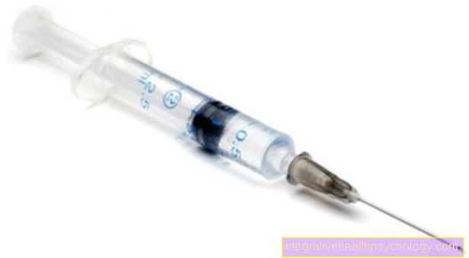
After disinfection, the affected area is numbed locally. For this purpose, local anesthetic is applied around the later entry point with a syringe. After a few minutes you are numb at this point. In the following, the correct process of skin biopsy is carried out and the skin sample is taken.
This can be done depending on the type of biopsy. In a punch biopsy, a small part of the cylinder skin is traumatically removed with a special device. With some biopsies, a piece of skin is cut out of the surrounding tissue with the help of a scalpel.
Then the edges of the wound are sewn together so that healing takes place better and faster. It also reduces the risk of infection. A plaster or bandage is then applied and the biopsy is complete.
What is a skin punch?
A skin punch is an apparatus that can be used to perform a punch biopsy. This apparatus has a cavity in the shape of a cylinder at the tip. This tip is used to penetrate the skin and take a cylindrical sample.
The tip leaves a lesion two to three millimeters in diameter. A skin punch is used when the skin change being examined is small. This results in fewer skin injuries compared to other methods.
How painful is that?
The skin biopsy is not painful if performed correctly. However, there may be pain throughout the biopsy process. Usually the beginning of the local anesthetic is the most painful moment. A little liquid local anesthetic is injected into the skin with a syringe and needle.
The puncture is often described as slightly uncomfortable and painful. It can also cause a burning sensation under the skin. Pain may also occur after the biopsy. This affects exactly the place where the skin was removed. This is like a small abrasion. However, since the affected area is very small, the pain is often not present here.
evaluation
The evaluation of a skin biopsy can be done quickly or only be available after a few days. Usually the skin sample is placed in a solution and sent to a special facility. The final evaluation then takes place here.
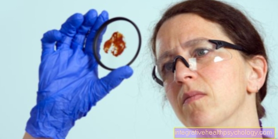
For the evaluation, the sample can be prepared so that it can be viewed under the microscope. It is either done by a dermatologist or a pathologist. The density of the skin is also often measured in order to be able to make further statements.
The attending physician will then be informed of the result of the skin biopsy. It is more rarely possible that the sample is evaluated on site after the biopsy has been taken. For this, however, high costs have to be raised for the necessary devices. Therefore, this is not to be expected in small doctor's offices or small hospitals.
Risks
There are some risks associated with removing skin during a skin biopsy. Usually, however, there are no complications because the wound that is created by the biopsy is very small. Nevertheless, this invasive measure can lead to bleeding. The bleeding is not very heavy and should stop on its own after a while.
A bruise can also develop in the area of the biopsy (see also: Hematoma) come. This will last for some time. Since the skin is a barrier, the risk of infection is also increased, as with an injury. A local infection of bacteria can occur or, in rare cases, an infection of the bloodstream.
You can also show a reaction to the local anesthetic. This is due to an intolerance and can be caused by redness or symptoms such as an allergy. The risk of a reaction to the local anesthetic is also low.
How quickly does the biopsy wound heal?
In general, the biopsy wound should heal as quickly as an abrasion. This means that within a few days of a punch biopsy, a crust should have formed. However, it can take up to two weeks for the wound to heal completely.
It can also happen that the miraculous edges are sewn together by the doctor during a skin biopsy. If self-absorbable sutures are not used, they should be removed within seven to ten days of the biopsy. This is followed by further healing within up to two weeks, in which the areas where the threads were attached heal.
This article could also be of interest to you at this point: Phases of wound healing
Duration
The duration of a skin biopsy can vary widely. How long a skin biopsy takes is also a matter of definition. As a rule, however, this only refers to the actual duration from the beginning of the action on the patient to the removal and subsequent bandaging of the wound.
If there are no complications during this time, the entire process is over within a few minutes. In some cases it can take up to ten minutes. With a preliminary examination, discussion, clarification, preparation of the doctor and sending in the samples, several weeks can pass from the first visit to the doctor until a final statement about the findings of the skin biopsy can be formulated.
costs
The cost of a skin biopsy can vary depending on the context. The type of biopsy does not change the costs. If the skin biopsy is performed together with another procedure or as part of a different disease, it will be billed differently than if it is performed alone.
As a rule, there are no separate costs for the patient. In addition, if complications occur, further measures that were carried out as part of the skin biopsy can be billed separately. For a skin biopsy, an amount of a few euros is usually charged. The laboratory costs around € 10.
What are the alternatives?
There are several other examination methods in dermatology. However, not a single one of these examinations can be used as an alternative to a skin biopsy, since only they allow a histological or cytological analysis of the skin and its cells.
With the other examination methods it is only possible to subjectively assess the cells with a much lower resolution. The advantage of these methods, however, is that they are less invasive. This means that no injury has to be inflicted on the patient in order to obtain a more extensive diagnosis.
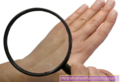
Some of these alternatives are an examination using ultrasound, a magnifying glass for magnification and follow-up monitoring to assess any changes. The assessment of the odor can also be an important aid in dermatology.