Medial meniscus
Synonyms in a broader sense
Cartilage disc, anterior horn, pars intermedia, posterior horn, inner meniscus, outer meniscus,
English: meniscus
definition
The inner meniscus is - together with the outer meniscus - a part of the knee joint. It serves as a sliding and sliding bearing between the bones involved. Due to its anatomy, it is affected much more often than the external meniscus in (sports) injuries.

I would be happy to advise you!
Who am I?
My name is dr. Nicolas Gumpert. I am a specialist in orthopedics and the founder of .
Various television programs and print media report regularly about my work. On HR television you can see me every 6 weeks live on "Hallo Hessen".
But now enough is indicated ;-)
The knee joint is one of the joints with the greatest stress.
Therefore, the treatment of the knee joint (e.g. meniscus tear, cartilage damage, cruciate ligament damage, runner's knee, etc.) requires a lot of experience.
I treat a wide variety of knee diseases in a conservative way.
The aim of any treatment is treatment without surgery.
Which therapy achieves the best results in the long term can only be determined after looking at all of the information (Examination, X-ray, ultrasound, MRI, etc.) be assessed.
You can find me in:
- Lumedis - your orthopedic surgeon
Kaiserstrasse 14
60311 Frankfurt am Main
Directly to the online appointment arrangement
Unfortunately, it is currently only possible to make an appointment with private health insurers. I hope for your understanding!
Further information about myself can be found at Dr. Nicolas Gumpert
Illustration of the medial meniscus
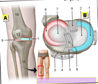
- Inner meniscus -
Meniscus medialis - Inner articular knot
(Shinb.) -
Medial condyle - Transverse ligament of the knee joint -
Lig. Transversum genus - Kneecap ligament -
Ligamentum patellae - Bursa - Bursa
- Outer meniscus -
Lateral meniscus - Outer joint nodules
(Shinb.) -
Lateral condyle - Anterior cruciate ligament -
Lig. Cruciatum anterius - Posterior cruciate ligament -
Ligamentum cruciatum posterius - Femur - Femur
- Shin - Tibia
- Kneecap - patella
You can find an overview of all Dr-Gumpert images at: medical illustrations
Anatomy and function of the menisci
The menisci of the knee are made of fibrous cartilage. They lie within the knee joint, which is formed from the articular knots (condyles) of the thighbones (femur) and shinbones (tibia) and the kneecap (patella). When viewed from the front, they lie like two wedges in the knee joint, with the base on the outside and narrowing towards the inside. Viewed from above, they are roughly C-shaped.
Due to the relatively small contact area between the upper and lower leg bones, which do not fit together perfectly (they are therefore incongruent), the menisci are necessary to optimize the articulation (the interaction) of these two surfaces and to optimally distribute the strong forces that act here . So they lie as a kind of "joint socket" on the tibial condyles (articular knots of the tibia). When the knee is flexed, they slide back up to one centimeter and return to their starting position when extended. This strong tensile and compressive load explains the susceptibility (at least of the inner meniscus) to injuries.
Anatomy and function of the medial meniscus
The medial meniscus is relatively precise C-shaped and greater as the External meniscus. It is at its front and rear ends (Front horn and Back horn) in the bone, in the so-called Intercondylar area (Area between the articular knots), anchored.
He's also sideways with the Joint capsule grown together. In addition to this, the inner meniscus is also connected to the Inner band (medial collateral ligament or collateral ligament, lig. collaterale tibiale) fused. Through this fixation he is in his mobility limited than the outer meniscus and thus comes under greater tension when the knee is moved. The inner meniscus becomes strongest at External rotation of the bent knee.
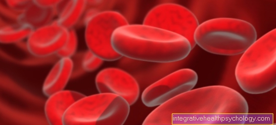
Blood supply
Both Menisci (Inner meniscus and outer meniscus) are not interspersed with blood vessels at all in their central part and only sparsely further outward. Therefore the outer - the area with the best blood supply also the name "red zone". The supply of the inner parts of the meniscus with nutrients happens mainly through the Joint capsule and the Synovial fluid (Synovia).
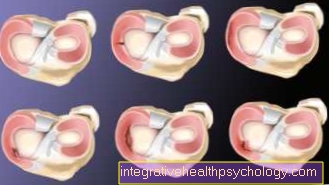
The poor blood supply causes that Lesions The menisci are slow to heal (damage). Ever further inside the damage lies, all the more worse the healing is taking place. This is important in the care of Meniscal tears, because cracks in the outer zone due to the better blood supply basically means seam can be treated. This is less possible in the case of damage to the inner part of the meniscus; here it is more likely to be partial distance indicated by meniscus tissue.
Basically, meniscus tissue should only be removed if a suture is not possible, this is explained by the fact that Osteoarthritis risk of Knee joint the less meniscal tissue remains, the higher it is.
Clinical significance
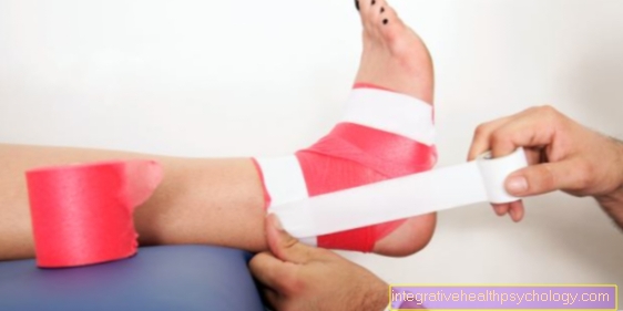
Meniscus lesions are among the most common injuries to the Knee joint. The inner meniscus is - due to its adhesion with the medial collateral ligament - essential more often affected by injuries than the External meniscus. Typically it cracks Rotational movements with bent knees, where the foot is fixed to the floor and does not turn with it.
For example with To ski or at Soccer game with studded shoes. Often it is not affected alone, but tears in the context of the so-called "unhappy triad“(Unfortunate triad), being an additional Tear of the medial collateral ligament and des anterior cruciate ligament (Ligamentum cruciatus anterius) are present.
Medial meniscus tear or damage to the medial meniscus
The median meniscus becomes acute most often in the context of Sports injuries damaged. Especially movements in the Knee joint with abrupt Stops, Rotational movements or Contortions can cause damage to the menisci. Sports in which such movements occur particularly frequently are for example Soccer, basketball, Tenniss and To ski. The most common non-traumatic cause of medial meniscus damage are wear-related Meniscal injuries. Through the wear and tear of the articular surfaces over the years or with constant Improper loading it comes to one Abrasion the menisci.As a result, they are already damaged on the one hand, and on the other hand become each other at the same time more vulnerable for traumatic injuries. A person with a worn meniscus will recover more quickly from exercise Meniscal tear to contract as a person with intact menisci. Incorrect strains that damage the menisci are, for example, congenital leg deformities (Knock knees or If one), as well as frequent work in the squat or Obesity. In the case of a degenerative process on the medial meniscus, the affected person usually feels under burden increasing pain in the knee. The extent of the pain differs depending on the severity of the injury.
When the meniscus is merely slightly torn the pain may be mild. The knee can often tear with a meniscus no more be moved within the normal range of motion. If parts of the meniscus have already rubbed off or lifted off, the knee can be bent or extended cracking noises trigger in the knee joint. When a meniscus in an accident suddenly tears, this is usually expressed by severe stabbing pain shooting into the kneethat make further strain on the knee impossible. In addition, it often develops Joint effusionwho become a swelling of the knee. A normal range of motion is then no longer given.
Part of the cartilage that has been blown off can also become acute blockade of the joint, which can then no longer or hardly be bent or stretched. The main medical tool for diagnosing a Internal meniscus tear is the physical examination. Doctors can use various orthopedic tests to check whether the menisci are involved. There will be different Handles, Pressure points and Movement sequences used and checked for pain. If the medial meniscus is damaged, the pain is concentrated in the inside of the knee joint gap. In imaging, the Magnetic resonance imaging (MRI of the knee) suitable to detect a meniscus tear. Also one Jointoscopy (Arthroscopy) can be used to assess the extent of meniscus damage.
This can be chosen for that Therapy procedure matter. The therapy of the meniscus tear is important, otherwise Long term complications can result. Especially if parts of the medial meniscus are already in the Joint space should have reached a operational Treatment of the injury should be chosen, as the free piece of cartilage due to the friction leads to another damage of the articular surfaces. In the long run, this in turn leads to a Osteoarthritis development in the knee joint. Depending on the location of the crack, the Meniscus suture to prefer. However, this can only be done in places where the inner meniscus is better supplied with blood is. Otherwise the seam will not be able to lead to the meniscus parts growing together. As alternative procedures come to such poorly perfused Areas the Partial meniscal resection or the complete meniscus resection in question. The partial resection of the meniscus is as little as possible Meniscal tissue away. Particularly free fragments are taken out of the joint space so that they do not lead to further damage to the joint surfaces. If the meniscus tear is very large, the entire meniscus must sometimes be removed in order to achieve an optimal therapy result. The removed inner meniscus must then pass through a graft or one artificial Meniscus to be replaced. Physiotherapeutic exercises to restore the mobility of the joint are particularly important. Depending on the extent of the injury, it can Weeks to months take until the knee is loaded normally again and again Sports may be driven. The attending physician must discuss this with the patient individually. Minor meniscus injuries do not necessarily have to be operated on. In this case, relieving the affected leg by immobilizing the knee for a few weeks, taking painkillers and physiotherapy often help to achieve good healing.
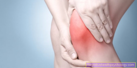
Medial meniscus pain
Injury to the medial meniscus can be very painful. Meniscus tears that occur suddenly, for example as a result of an accident or a sports injury, usually lead to severe pain in the affected knee joint. If a piece of cartilage loosens completely or protrudes into the joint space, this can lead to an abrupt blockage of the mobility of the knee. The pain that occurs in the menisci as a result of a degenerative process is more diffuse and less intense. They express themselves primarily in stressful situations and increase with the extent of the stress. Pain in the area of the joint space, which can also be triggered by pressure with the fingers, or pain when the knee is turned outwards, speak for damage to the inner meniscus (External rotation), as well as pain when squatting down or getting up from a crouched position. In addition, a joint effusion can develop as part of a meniscus injury. During this process, fluid collects in the joint space and presses on the surrounding structures. Depending on the extent of the effusion, it can also cause pain, as the knee is then very tight and tense. If a meniscus tear is not properly treated, osteoarthritis of the knee joint can develop over the long term. This is also characterized by pain when moving the knee. To prevent this, early therapy that is individually tailored to the injury is essential. Surgical intervention is not always necessary.
Posterior horn of the medial meniscus
The human knee has two meniscus - the outer meniscus and the inner meniscus. These form the Articular surface and can be further divided into different parts. The medial meniscus, which lies on the inner side of the knee joint, also has a part called the posterior horn. This is the part that does the rear Articular surface on the inner Side of the knee joint. At Lesions of the medial meniscus, the medial meniscus posterior horn is special frequently affected. Depending on the extent of the crack, the patient feels different degrees of intensity Pain, can no longer move the knee properly and develops joint effusion. Treatment of a tear in the posterior horn of the medial meniscus depends on the severity of the injury. Basically, a conservative Therapy take place. However, if the cartilage has loosened or the tear is very large, then it is operational Preferred care for the injury.


.jpg)