Head hematoma in the baby
What is a cephalic hematoma?
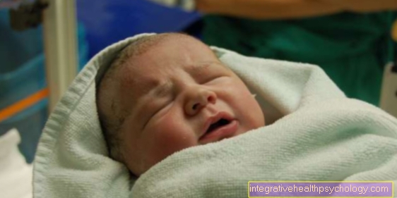
The cephalic hematoma, also known as a "head blood lump", is a bruise that occurs in connection with an injury to the baby at birth. This results in vascular injuries to the back of the child's head as a result of shear forces during the birth process.
The cephalic hematoma is defined as such by the fact that it is located directly above the skull bone and below the associated periosteum, which gives it a typical elastic consistency. This location also prevents the bruise from expanding beyond the limits of the skull bone and defines the cephalic hematoma
causes
The cephalic hematoma is caused by shear forces between the bones of the skull, which typically arise during childbirth. In this context, so-called “seam diastases”, that is, the skull bones slipping apart, and skull fractures often occur.
In both cases, this often injures vessels between the skull bones and between the bones and periosteum. The resulting bleeding is deposited between the bone itself and its periosteum and causes a bulging, elastic bruise that does not spread beyond the boundaries of the affected skull bone.
Such hematomas are often caused by a so-called "Forceps delivery“Contributed. This is a delivery that should be facilitated with the help of forceps. Uneven pressure is exerted on the child's head, which promotes a cephalic hematoma.
diagnosis
The diagnosis results to a large extent from the characteristics of the cephalhematoma and how it presents itself. This includes its firm, elastic consistency, which is due to the fact that blood collects between the skull bone and its tight periosteum. In addition, this also results in its spreading pattern, which does not extend beyond the boundaries of the skull bone.
In addition, sonography is used to diagnose the cephalhematoma. It should provide information about where exactly the bruise is and where it is expanding. The brain and skull bones are also assessed in order to rule out further injuries.
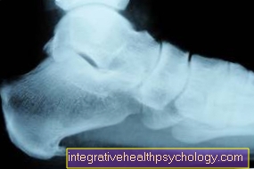
However, if there is a suspicion of a fracture of the skull, further imaging methods are required, such as an MRI, in order to better illustrate the possible fracture.
Concomitant symptoms
Other birth injuries, such as skull fractures or other head tumors, are often associated with a cephalic hematoma. These include the "Caput succedaneum“, Which is also called a birth tumor and consists of fluid located under the skin. It completely regresses within a few hours to days without any additional action.
The "subgaleatic hematoma“Consists of a bruise that sits over the periosteum and is also caused by shear forces. During childbirth, the child's collarbone can also break or various paralysis caused by nerve irritation or squeezing. They, as well as the fractures and tumors, heal largely without consequences.
Read more on the subject: Broken bones in the baby
treatment
In the treatment of a cephalhematoma, proper prophylactic administration of vitamin K should be ensured. A vitamin K deficiency leads to a disruption of the coagulation and can cause or increase bleeding. In general, you can only wait for the body to break down the bruise, which can take weeks to months, depending on the size of the tumor.
If the findings are very pronounced, however, a relieving puncture of the bruise can provide relief, but carries a serious increasing risk of inflammation and should therefore be carefully considered. It looks different if there is an additional fracture of the skull that does not follow a straight line or whose fracture components are indented. In this case, a surgeon will need to treat the hernia and remove the bruise as much as possible.
You might also be interested in the topic: U2 examination-vitamin K administration
When is an operation necessary?
An operation only needs to be performed if an additional skull fracture has occurred that does not follow a straight line or the fracture components have been dented. To correct this condition, a surgeon must treat the fracture site and allow the skull bones to grow together properly. The bruise can also be removed, which facilitates the healing of the cephalhematoma.
In addition, an operation is of course necessary if the brain or other organs have suffered a birth injury. In conclusion, one can say that no surgery is necessary to treat a cephalhematoma, provided that no complications or further injuries have occurred.
Can Osteopathy Help?
I would be more careful here, as the cephalic hematoma is a bruise that is due to shear forces on the skull. This means that further manipulation can lead to further bruising, since the infant's skull is not completely fused and therefore offers little stability.
In individual cases, however, it can be discussed with the osteopath what he thinks is helpful and does not harm the baby.
What are the long-term effects?
One of the long-term consequences of a cephalhematoma is ossification of the edge of the hematoma in the course of healing. This leads to hardening at the edge of the bruise while a soft depression develops in the middle.
At first glance, this property makes it easy to mistake the healing bruise for an indented fracture of the skull.
Duration
In most cases, the cephalic hematoma resolves within a few weeks to months without further action. This course always depends on the size of the bruise, as well as the fact whether there are other injuries and whether the coagulation of the infant is healthy. The healing of the bruise should be checked by a doctor from time to time.
What complications can arise?
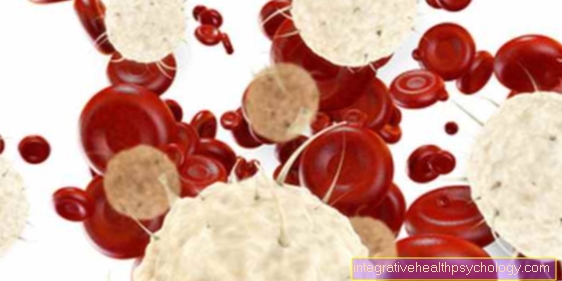
The fact that a not insignificant amount of blood flows into the hematoma results in anemia in some cases which must be treated. A massive head hematoma can trigger a so-called "shock", which can be life-threatening because not enough blood is allowed to circulate. With such pronounced bruises, especially in male newborns, it should also be examined whether a coagulation disorder is partly responsible.
In addition, it must be checked that there is no iron deficiency due to blood loss, which in turn can lead to anemia. Newborn jaundice can occur due to the breakdown of the stored blood in the hematoma. This is another reason why a cephalhematoma should be examined by a doctor and re-examined from time to time.
Read more on the topic: Newborn jaundice
What is a calcified cephalic hematoma?
A calcified cephalic hematoma is the process of breaking down the bruise. As part of the healing process, ossification can occur in the edge area and a soft depression in the middle. At first glance, this stage can easily be confused with a dented skull fracture. As the healing process progresses, these calcifications dissolve and the cephalic hematoma usually heals without any consequences.