Collagen
Layout and function
Collagen is a protein which, as a structural protein, makes up an essential part of connective and supporting tissue. Accordingly, it occurs in most of the organs in our body. Collagen is one of the fiber proteins and has a specific anatomical structure so that it results in a stable protein. The collagen molecule has a basic structure made up of three polypeptide chains. These are strung together proteins that consist of up to 1000 individual amino acids, the smallest unit of proteins.
The synthesis of a precursor to collagen is first produced in the cells. The three protein chains lie together and twisted around each other. They form a clockwise rotating basic structure, which results in a collagen molecule approx. 300 nm long and 1.5 nm thick. This arrangement is known as the triple helix and forms the precursor of collagen. The further production of the collagen now takes place outside the cell. Certain enzymes cut off peptides from this procollagen at the ends. Now the individual triple helices can be arranged in parallel and form cross bridges. This means that the colleague molecules network with each other and thus form a stable and associated framework.
In the light microscope, you can see a typical horizontal striation, which is caused by the fact that the colleagues' molecules lay together to form fibrils and the ends overlap. Several fibrils eventually form a collagen fiber. Then water molecules bind to the finished collagen, which means that collagen always has a high water content. The assembly of different peptide chains creates different triple helices. Therefore, a distinction is made between different types of collagen, which are usually numbered consecutively, such as collagen type 1, type 2 or type 3.
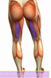
The types of collagen occur differently in the different types of tissue in our body. In general, collagen is found in skin, bones, fiber cartilage, tendons, ligaments, teeth, muscle skin and in the eyes. The contained collagen gives these structures the necessary strength and stability. Due to their very elastic properties, bones, cartilage and tendons are tear-resistant, but also flexible.
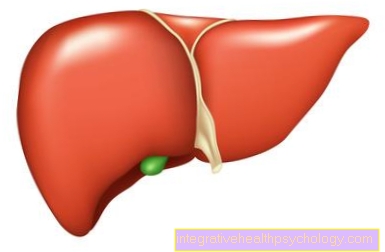
In bones and teeth, it is also involved in mineralization, the build-up of bones and tooth enamel, and is an important part of the metabolism there. The organs of our body are usually enclosed by a capsule and fatty tissue. Collagen also forms surface layers and is mostly located in the connective tissue. The organs are thereby separated from each other, but remain flexible in their position. Collagen is therefore also involved in the cushioning and elasticity of our organs and takes on a protective function with the fatty tissue.

Collagen in the skin
A very large proportion of collagen is in the skin, where it takes on an important support function for the skin layers and the adjacent connective tissue. As a protein, collagen has the property of binding water, which keeps the skin taut. Due to the special structure of the collagen, the collagens are very elastic, which means that the skin is also very elastic and flexible. How important collagen is to that Skin firmness is shown when the collagen content slowly decreases from around mid-20s.
Little by little, the first wrinkles appear, which has to do with the breakdown of collagen in the skin. The skin then loses its elasticity and collapses. The own collagen production decreases significantly, which is why different ones Cosmetic products in the form of creams or collagen building blocks such as proteins and amino acids try to fill the collagen cushion from the outside. Collagen-containing creams or direct injections into the skin should smooth the wrinkles again and make the skin appear tighter. Since the collagen binds water, the skin should look firmer and fresher again immediately after an injection treatment.
Types of collagen
Type 1
When it comes to collagen, a distinction is made between different types, each of which has a different proportion in different organs. Type I collagen is approx. 300 nm long and forms the typical structure of densely packed collagen fibrils, which can be between 50 and 200 nm thick. In terms of quantity, type 1 collagen is the most common in the human body. This type is particularly common in the skin, connective tissue, tendons, bones, muscle fascia and the cornea. In the structures mentioned, the collagen is in the extracellular matrix, which means that the collagen surrounds the individual cells in the skin, bones and tendons.
By storing water in the collagen, the organs gain mechanical strength. The high collagen type 1 content in the skin and tendons makes them particularly strong and flexible. The proportion of colleagues ensures the necessary compressive strength and firmness of the various structures.
One of the most well-known disorders of collagen type 1 synthesis is the Osteogenesis imperfecta. This is the Vitreous bone disease, a hereditary defect in bone formation. As a result, too little collagen is produced and the bone is less stable and resilient. Depending on the severity of the disease, this can be different. Step at the patient spontaneous and frequent fractures on. Deformities of the skull and spine can also occur. In addition, patients usually do not grow very tall, as the disease affects all bone growth.
Type 2
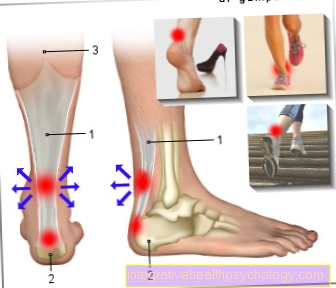
Type 2 collagen, like type 1, is also a fibril-forming collagen. In terms of length, the two types are very similar. Type 2 is about 300nm long, but it is usually thinner than type 1 collagen. Type 2 collagen is particularly common in hyaline and elastic cartilage in front. Hyaline cartilage lines the joints of the body and forms the top layer of the joint space.
Elastic cartilage occurs, for example, on the auricle, the ear canal and also in the small bronchi of the lungs. While type 1 collagen has a dense structure, type 2 collagen fibers lie loosely in the various structures in the connective tissue. In addition to the collagen there are other substances such as proteoglycan and Hyaluronic acid in the cartilage. Due to this composition and accumulation of water, the cartilage becomes pressure-resistant, elastic and stretchable, but is not as stable as bones.
Hydrolyzate
Hydrolysates are products that result from the breakdown of proteins or proteins. Hydrolyzate can also be obtained from collagen by enzymatic cleavage (hydrolysis). These collagen proteins are preferably obtained from type 1 collagen and used as Food supplements utilized. They contain a high proportion of short amino acid chains (peptides) and are very similar to gelatine.
One difference is that collagen hydrolysates do not gel and can be easily dissolved in water. It is a white, odorless and tasteless powder that can be used for binding, emulsifying and foaming. This powder is used especially in protein-rich diets and the Sports nutrition. It is available as a powder that can be dissolved and supplements the protein intake during vigorous physical activity. It is also used to repair damaged cartilage tissue.
The collagen hydrolysates are supposed to stimulate cartilage formation and thus regenerate worn joint material. In patients with cartilage wear (osteoarthritis), this should lead to an improvement in pain and mobility of the joints. The collagen proteins are also contained in some cosmetic products, since they are well absorbed by the body, they should be able to penetrate into the deeper layers of the skin and improve and tighten the appearance of the skin.