windpipe
Synonyms
Lat. = Trachea; Function of the trachea, anatomy of the trachea
English: windpipe
definition
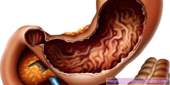
The trachea, along with the bronchi and lungs, are part of the lower airways and connect the nasopharynx with the lungs. The windpipe can be found in the throat below the larynx (larynx) and in the chest (thorax).
Illustration of the trachea
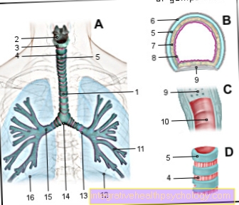
- Windpipe (approx. 20 cm) -
Trachea - Thyroid cartilage -
Cartilago thyroidea - Cricoid cartilage -
Cartilago cricoidea - Ring band -
Annular ligament - Tracheal cartilage -
Cartilago trachealis - Cover fabric - Tunica adventitia
- Trachea glands -
Glandulae tracheales - Mucous membrane - Tunica mucosa
- Membrane rear wall -
Pariesmembranaceus - Tracheal Muscle -
Tracheal muscle - Bronchiole - Bronchiolus
- Left lung -
Pulmo sinister - Left main bronchus -
Bronchus principalis sinister - Bifurcation of the windpipe -
Bifurcatio tracheae - Right main bronchus -
Bronchus principalis dexter - Right lung -
Pulmodexter
You can find an overview of all Dr-Gumpert images at: medical illustrations
Anatomy trachea
The air you breathe makes its way from the Nasal cavity over throat (Pharynx) and Larynx (larynx) to windpipe (Trachea) and from there to the Bronchi of the lungs.
The trachea is a 10 to 12 cm long, elastic tube with a diameter of 12 mm. It is divided into two sections that Pars cervicalis („Neck part“) As well as the Pars thoracica („Rib cage part“).
Based on the location of those behind it Spine the windpipe begins at the level of the 6th / 7th Cervical vertebra and ends at the level of the 4th Thoracic vertebra. There it divides into the right and left Main bronchus of the lungs and forms a bifurcation at this point (Bifurcatio tracheae, "Branch") with a cartilaginous groin (Carina tracheae).
The trachea is made up of 10 to 20 horseshoe-shaped cartilage braces, which are connected to one another by ligaments in the longitudinal direction Annular ligament (Ligament = Band, annulus = Ring) are connected.
Histology / tissue
The histological one Structure of the trachea is three-layered (from inside to outside):
- Tunica mucosa = Mucous membrane with glands
- Tunica fibromusculocartilaginea = Musculature, Cartilage, ligaments
- Tunica adventitia = surrounding connective tissue
The tunica mucosa consists of a multi-row ciliated epithelium, which is covered with cilia, so-called Kinociliais busy. Mucus-producing goblet cells are stored. You can also find Support cells, Basal cells such as endocrine cells.
The demarcation to the underlying Tunica fibromusculocartilaginea forms a layer of connective tissue with elastic fibers and glands, the glandulae tracheales (Glandula = Gland).
The middle part of the windpipe consists of cartilage clips that are open to the rear hyaline cartilage. The ends of a clasp are connected by means of a Muscle tendon plate (Tracheal muscle), which forms the back wall of the windpipe. Between two cartilage braces there is one connective tissue flexible connection (Annular ligament).
Finally, the outermost layer that Tunica adventitia, forms loose connective tissue and anchors the windpipe in its surroundings.
function
The windpipe as part of the air-conducting (conductive) Airway serves the warming, Moistening and cleaning the breath.
This is done with the help of the mucus producing goblet cells as well as the Kinocilia the mucous membrane. The latter transport slime and foreign particles at a speed of about 15 mm per minute towards the throat.
One can find more Nerve fibers in the trachea, which is responsible for the Cough reflex are responsible and in this way also take on a cleaning function.
Trachea pain
Trachea pain can have various causes.
The most common reasons are Inflammation of the airways. If there is pain in the area of the windpipe, the inflammation is most likely in the area of the Throat, Larynx or in upper trachea.
Come as a pathogen Viruses, bacteria and also with immunocompromised people Mushrooms in question. Smoking favors such infections, as the mucous membrane that lines the airways is damaged and the penetration of the pathogens is made easier.
The pain in the windpipe is often seen as a burning sensation in and behind the throat Sternum felt. In principle, however, anything that can injure and irritate the lining of the larynx and windpipe can cause pain.
This also applies to inhalation of toxic chemical irritant gases, such as pepper spray. In patients with a Reflux disease, better known as heartburn, it is possible that not only the esophagus but also the windpipe can be affected.
Especially with severe illness and when lying down, for example while sleeping, the acidic gastric juice can along the esophagus up into the throat, from there into the windpipe and attacking its mucous membrane.
The consequences are also pain and the affected vocal cords hoarseness.
Even completely healthy people can experience pain in the windpipe and lungs. Everyone knows it during physical exertion in winter in cold air. The cold air is a minor inflammatory stimulus that affects the lining of the airways.In spite of all this, physical stress at temperatures greater than minus 15 degrees is harmless for otherwise healthy people and the advantages outweigh the disadvantageous inflammatory stimulus.
Asthmatics should be more careful when exercising in winter temperatures, as the lungs are already under chronic inflammation due to the disease and an additional stimulus can cause an asthma attack, which is also associated with pain, but especially in the chest area.
Burning sensation in the windpipe (tracheitis)
There is often a burning pain in the windpipe behind it Inflammation of the trachea.
In medical parlance it is called Tracheitis designated. It is a rare disease that often arises as a result of a pre-existing cold infection.
The Tracheal lining is pre-damaged by the cold viruses, more rarely bacteria, which in turn allows bacteria to penetrate them and infect the windpipe.
However, the cold viruses are also able to descend from the nose and throat into the windpipe and infect them directly. The disease occurs mainly in the winter months.
Usually, trachitis does not occur in isolation, but in combination with one inflamed larynx (laryngitis) and inflamed bronchi (bronchitis) on. In addition to an infectious cause, irritant gases, for example, can lead to a searing and painful inflammation of the windpipe.
Tracheitis is a rare disease and complication of a common cold, but it should definitely be treated by a doctor. If symptoms such as cough, hoarseness and burning sensation behind the breastbone persist for a few days, the use of an antibiotic is recommended.
The tracheotomy
Under a Tracheal incision one describes an artificially created opening of the windpipe.
A type of tube / cannula is then inserted into this opening, which connects the trachea with the outside world and keeps the incision open. This tube, which carries the air through the cut in the windpipe into the Lungs conducts, is used in medical parlance as "Tracheostomy" designated.
stoma is the encroachment for an artificially created body opening.
A tracheotomy is called a Tracheotomy designated. The tracheotomy becomes necessary when a person is no longer able to breathe independently and is dependent on external ventilation, for example from a machine, for a long period of time.
This is especially the case with patients in the coma the case.
Even in patients with Throat cancerwhich obstructs the airflow in the windpipe and lungs or has made it necessary to remove the larynx, often require a tracheostoma.
The term Tracheal incision is often used incorrectly in common parlance. Many imagine the incision made in the throat in acute, life-threatening shortness of breath.
This "Emergency tracheotomy“Is correctly called Cricothyrotomy where the larynx and not the windpipe is incised. While the cricothyrotomy is an emergency medical procedure if there is a risk of suffocation, the tracheotomy is planned to be carried out when the patient is in a foreseeable long-term ventilation situation.
Complications of a tracheal incision are injuries to the larynx, the thyroid gland or those located behind the windpipe esophagus Bleeding and infections, especially if the tracheostomy tube is used for a long time.
Tracheotomy
A so-called Tracheotomy can be carried out if, for example, long-term ventilation is required. Here, a cannula with a connected ventilation hose is inserted into the upper part of the trachea between the 3rd and 4th cartilage brace, so that air can flow in over it and the oxygen supply to the lung is guaranteed in this way. The resulting hole is called a tracheostoma (stoma = mouth, opening).


.jpg)