MRI of the hip
General
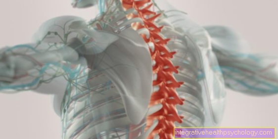
Magnetic resonance tomography (MRT) is an imaging technique that can be used to show particularly soft tissues. In contrast to x-rays or computed tomography, the patient is not exposed to any radiation. The images are created using magnetic fields and radio waves that align certain particles of the body in one direction. When the magnetic field is switched off, the particles orient themselves again in their original orientation and the time until they have reached this is measured. The resulting images are then based on these measured values.
Soft tissues such as ligaments, tendons, muscles and blood vessels can be seen particularly well on MRI images. Cartilage is also very visible. Just like water or other fluids that are in the joint.

pool
The pool or that hip joint occupies a central position in the human body, as it is exposed to a high level of stress through constant standing and walking. With a lot of movements it has to carry a multiple of the body weight and has to guarantee a high mobility.
Because of this high load, the hip joint is often affected by injuries or shows signs of wear and tear, which with the help of the MRI can be proven.
A MRI of the pelvis and an MRI of the hip is an MRI of the same region. Depending on the question, a special focus is placed on the pelvis or the right or left hip.
You can find more information under our topic: MRI of the pelvis
application areas
Since the MRI scan provides much more detailed images than a simple X-ray, it is not only used to clarify bone fractures, but also to detect or exclude diseases of the soft tissues of the joint.
In addition to bone fractures, for example a fracture of the femoral neck, a dislocated hip (hip joint dislocation) can also be detected on the MRI of the hip.
Injuries to the joint capsule, such as a tear off of the capsule from the so-called joint lip (labrum tear), are often shown well on the MRI images. Inflammation of muscles, ligaments or bursa can be recognized as well as cracks in structures. Magnetic resonance imaging (MRI) of the hip also provides information about the progression of degenerative bone diseases such as hip arthrosis or femoral head necrosis. Due to the precise representation, these can be diagnosed at an early stage.
Other joints such as the sacroiliac joint on the rear hip can also be checked for osteoarthritis with an MRI.
Read our article about a MRI of the sacroiliac joint
In contrast to X-rays, water retention in the joint (edema) can also be shown in an MRI examination.
An MRI of the hip is very important in the area of tumor diagnostics, in the area of the hips especially for the representation of tumors in the femoral neck.
preparation
An MRI scan of the hip does not usually require any special preparation. It will be a Educational talk with the doctor, where all possible questions can be answered. In addition, the doctor should discuss any intolerances Contrast media be informed. Also any existing ones Claustrophobia should be communicated to the doctor in order to consider whether a Sedatives should be given.
If you have a pronounced claustrophobia, you can also use our MRI for claustrophobia topic to find out about the possibilities that exist in the case of claustrophobia anyway to perform an MRI of the hip.
Before starting the investigation, you must all metal-containing objects away from the body become. This applies to jewelry, piercings and metal that is on clothing, such as underwired bras, trouser buttons, etc., and chip cards such as debit cards, purses and keys may not be taken into the examination room because they are affected by the strong magnetic field attracted and thus damage the patient and the MRI machine. The same applies to all electronic devices, such as cell phones or MP3 players.
Contraindications
In patients who have a built-in Pacemaker or an implantable one Defibrillator (ICD), magnetic resonance imaging is usually not allowed, as both the patient and the device can be damaged.
For some time now there have also been pacemakers that can withstand the examination; this should be clarified with the doctor in advance. Also used metal parts, such as wires in bones or artificial ones mechanical heart valvesshould be reported to the doctor. He must then decide whether the examination can be carried out.
Also an artificial inner ear (Cochlear implant) is a contraindication to an MRI scan of the hip.
You can find out more about the topic here: MRI and implants
With a known functional restriction of the kidney Magnetic resonance imaging can be performed, but the administration of Contrast media be waived.
Procedure for an MRI of the hip
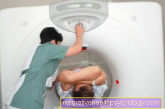
When all metal parts have been put down, the MRI examination to be started. To do this, the patient lies down on a bed that is in the MRI tube can be driven in.
For an MRI of the hip, the patient is shown with the head moved forward into the tube until the hip is also in the tube. People who suffer from claustrophobia should report this to the doctor beforehand, so that he can prescribe a sedative if necessary.
Before the patient is driven into the tube, he gets Soundproof headphones or ear plug with music on the ears.
These are intended to dampen the loud knocking noises that are generated by the device during the examination. In addition, the patient is given a switch that he can press if he wants to be pulled out for certain reasons or if he is not feeling well.
The radiology assistants are in an adjoining room, but can follow the examination through a pane of glass and see when the button is pressed by the patient. Both rooms are connected to each other via an intercom.
The MRI scan of the hip is after about 15 to 20 minutes completed.
If images are also taken with a contrast medium, this is injected after the first row has ended and a second exposure process follows.
Following the actual MRI examination, there is a final discussion with a radiologist who will evaluate the images.
Move out
Usually one has to sign up for an MRI scan Do not take off your hipsas the MRI images can also be made through clothing. Only the shoes should be taken off.
However, it is necessary take off all clothing containing metal.
These can be trousers or tops with metal buttons, or underwire bras. If you do not want to take off your clothes, you should make sure in advance that you are only wearing clothing without metal, such as leggings and a cotton shirt.
Complications
Since the Magnetic resonance imaging If there is no radiation exposure for the patient on the hip, complications or side effects are not to be expected if all preparatory safety measures (removing metal parts) are observed.
Tattoos or make-up on the skin can but that skin heat at this point by the magnetic rays and skin irritation can occur here.
Side effects may occur with the administration of Contrast media occur if this is not tolerated by the patient. Then it can become a allergic reaction come.
At pregnant women Women can experience complications during the first three months of pregnancy. Therefore, in this case, an MRI should be avoided if possible if there is no emergency.
Please also read our topic on this MRI in pregnancy.
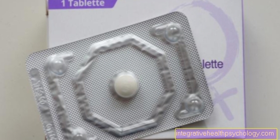
With contrast medium
By the gift of one Contrast media the effect is that different structures, which have quite similar gray levels in a normal MRT image, can be better delimited from one another. The contrast medium is in the vein injected and distributed in the Bloodstream. Vessels can therefore be seen much better, just like Tumors and Metastasesthat have a strong blood supply. An MRI with contrast agent is therefore usually more detailed and informative than an image without contrast agent.
With a special form of a Hip MRI, the so-called Arthro-MRI of the hip, the contrast agent is injected directly into the joint. Here, the smallest changes can be assessed more precisely than when contrast medium is administered into the vein.
As a contrast medium is mostly used Gadolinium GPDA used. It is a metal combined with an acid. MRI contrast media are generally well tolerated and can despite existing allergy against X-ray contrast media are used, as the means used in MRI no iodine contain. The contrast agent is excreted in the urine within 24 hours. Therefore, contrast medium administration should be avoided if the kidney is severely impaired.
In very rare cases the contrast medium can cause a so-called nephrogenic systemic fibrosis come. This is a connective tissue remodeling that, in addition to the skin can also affect the internal organs.
Without contrast media
MRI images without contrast agent can reveal major changes, but they are not suitable if an illness in the initial stage or the smallest changes or possible metastases are to be detected.
Your advantage of an MRI without contrast agent is that you have almost no side effects, because neither Radiation exposure, nor allergic reactions due to the contrast agent.
diagnosis
The MRI of the hip is now a very good imaging method to detect structural changes in the hip and other joints to be recognized at an early stage. Better than that roentgen and at one Computed Tomography can even change the smallest of bone and soft tissues are recognized. For example, a Hip arthrosis already in the initial stage by the injured party cartilage be recognized. Even small tumors or their metastases can be detected at an early stage by administering a contrast agent.
By very far advanced hip osteoarthritis MRI diagnostics can provide information about whether the hip can be preserved or whether a artificial hip must be used.
presentation
On the images of a magnetic resonance tomography, you can mainly see the soft parts of the joint, i.e. the joint capsule, Tapes, Tendons, Muscles and blood vessels. Cartilage is also clearly visible. Water or bruises in the joint can also be clearly seen. Above all, the administration of contrast media emphasizes the various structures very clearly and even small-sized tumors and metastases can be seen. These stand out either light or dark. It depends on how the pictures were taken
costs
The costs of the MRI recordings are usually covered by the statutory and private health insurance companies, as they represent a necessary diagnosis. Depending on the examination location and the effort (with or without contrast agent), the costs amount to approximately 400 to 1000 euros.
You can find more detailed information on this topic at: Cost of an MRI examination
Duration of an MRI scan of the hip
The MRI examination itself, i.e. the taking of the recordings, is about 15 to 20 minutesn not very time consuming.
It takes longer if you have additional recordings Contrast media be made. In addition, the preparation and waiting take a certain amount of time.
After the recordings, you have to wait for the doctor to conduct the final discussion with the evaluations of the images. The duration of the examination is difficult to predict and depends on the organization of the implementation.

.jpg)