Nerve cell
Synonyms
Brain, CNS (central nervous system), nerves, nerve fibers
Medical: Neuron, ganglion cell
Greek: Ganglion = node
English: nervous system
Also read:
- Nervous system
definition
Neurons (Neurons) are cells whose primary function is to transmit information with the help of electrical excitation and synaptic transmission is. The totality of nerve cells and other cells that are directly related to their function are called the nervous system, a distinction being made between the central nervous system (CNS), consisting of the brain and spinal cord, and the peripheral nervous system (PNS), mainly consisting of peripheral nerves.

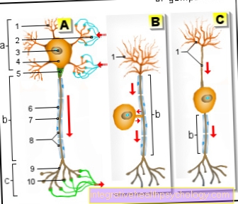
Illustration of a nerve cell
Nerve cell -
Neuron
- Dendrites
- Synapse
(axodendritic) - Cell nucleus -
Nucleolus - Cell bodies -
Nucleus - Axon mounds
- Myelin sheath
- Ranvier lace-up
- Swan cells
- Axon terminals
- Synapse
(axoaxonal)
A - multipolar neuron
B - pseudounipolar neuron
C - bipolar neuron
a - Soma
b - axon
c - synapses
You can find an overview of all Dr-Gumpert images at: medical illustrations
The human brain contains between 30 and 100 billion Neurons. Like other cells, the nerve cell has a nucleus and all other cell organelles that are in the cell body (Soma or Perikaryon) are localized.
A stimulus that hits a nerve cell causes an excitation that is in the Cell membrane of the neuron spreads (depolarization of the cell membrane) and over long cell extensions that Neurites or Axons, is forwarded.
This excitement is called Action potential. The neurites (axons) can reach a length of up to 100 cm. The excitation can thus be propagated over a long distance in a directional manner, e.g. when you move your big toe. Each nerve cell has only one axon.
construction
Nerve cells are divided into different parts. Every cell has a nucleus with a surrounding cytoplasm and cell organelles. This central area of the cell is called Soma. The Soma of the nerve cell has one or more thin processes that extend into Dendrites and Axon can be divided. Dendrites make contact with other nerve cells (synapses) and can passively transmit electrical excitation. If this excitation exceeds a certain threshold, an action potential is triggered in the axon voltage-dependent sodium channels open, which transmit this excitation over the entire length of the axon. In this way, a signal can be passed on over large distances within a short time. Axons can be over a meter long (e.g. motor fibers from the spinal cord to the foot muscles), so that excitatory nerve cells are among the largest cells in the body.
The axon either enters a single synapse to another nerve cell (e.g. in sensory nerves), or it branches out and makes contact with several cells (e.g. in nerves that innervate muscles). At these synapses in the cytoplasm of the cell are so-called. Transmitter vesicle before, small membrane-enveloped vesicles, which in high concentration messenger substances (Neurotransmitters) contain. If necessary, these can be released into the synaptic gap and trigger a signal on the cell membrane of the postsynapse - i.e. the target cell.
Nerve processes are made up of cytoskeletal elements such as the Microtubules streaked. These are tube-like protein building blocks that act like rails as a route for transport proteins (Dynein and Kinesin) which transport biological loads such as large proteins, vesicles and even entire cell organelles. In this way, the supply of distant axon elements can be ensured.
Many nerve cells are also surrounded by extensions of other cells in order to achieve better electrical properties (myelination). As a result, the nerve fibers increase in diameter, but can pass on excitation much faster. Motor fibers to skeletal muscles, for example, but also pain fibers, which are supposed to trigger a protective reaction, are particularly well covered.
You might also be interested in the following article: Structure of the nervous system
function
Nerve cells are able to process input signals and, based on this, pass on new signals. One distinguishes between excitatory and inhibitory nerve cells. Exciting nerve cells increase the likelihood of an action potential, while inhibitory ones reduce it. Whether or not a nerve cell excites depends on the neurotransmitter that this cell releases. Typical excitatory neurotransmitters are Glutamate and acetylcholine, while GABA and glycine inhibit. Other neurotransmitters like Dopamine can either excite or inhibit the target cell depending on the type of receptor. The stimulating and inhibiting signals that reach the nerve cells are integrated spatially and temporally and “converted” into action potentials.
A single signal that hits a nerve cell does not have to have any effect; in contrast to muscle cells, where every signal leads to the opening of ion channels and thus a contraction of the muscle cell. If, on the other hand, the excitation of the nerve cell is supra-threshold, this applies All-or-nothing principle: the triggered action potential always has the same amplitude. A modulation of the activity can only take place via the frequency of the action potentials, not via their intensity. The situation is different with signals that emanate from axons of other nerve cells: here, the cells can become more sensitive to this signal due to increased excitation over time. This phenomenon is called Long term potentiation and is jointly responsible for learning processes and memory formation, for example.
Functions of the nerve cell
As the eponymous cells of the nervous system, neurons are of vital importance Sensory, motor, coordination of vegetative functions and cognitive performance. The nervous system can be functionally divided: that somatic nervous system takes on tasks that are important for the interaction with the environment. This includes the innervation of skeletal muscles and the perception of external stimuli, for example via the sense of sight. The autonomic nervous system coordinates the function of internal organs and adapts their activity to environmental stimuli. It can be further divided into the sympathetic, parasympathetic and enteric nervous systems.
The sympathetic nervous system has functions which in the sense of a Fight-or-flight response, i.e. a stress reaction to environmental stimuli, are necessary. Heart strength and blood pressure are increased, the bronchi expand and the activity of the gastrointestinal tract is reduced. Conversely, activation of the Parasympathetic nervous system to an activation of the gastrointestinal tract (Rest and digest) and a decrease in blood pressure and heart work. The enteric nervous system, on the other hand, works primarily independently of the central nervous system and coordinates functions within the gastrointestinal tract and is modulated by the sympathetic and parasympathetic nervous system. The central nervous system can, however, be divided into core areas with motor, sensory, sympathetic, parasympathetic and higher cognitive functions that can be found in different locations of the brain or spinal cord.

Figure nerve cells
- Nerve cell
- dendrit
A nerve cell has many dendrites, which act as a kind of connecting cable to other nerve cells in order to communicate with them.
Read more about the topic here dendrit
Besides the neurites, which only lead in one direction, there are other processes on the nerve cell that Dendrites (= Greek tree). The dendrites are much shorter than the long neurite and are located near the cell body (perikaryon). Mostly they are in the form of a large dendrite tree in front.
Their job is to receive stimuli from other nerve cells. The connecting element, the "interface" between individual neurons is called Synapse.

Illustration of nerve endings / synapse
- Nerve ending (axon)
- Messenger substances, e.g. Dopamine
- other nerve endings (dentrite)
The end of the long nerve cell extension (axon end) of one neuron meets the dendrite tree of another neuron. The interaction between the two occurs through a chemical one Carrier substance, one Neurotransmitters; the process is similar to an "electrochemical coupling".
A nerve cell can be linked in this way with up to 10,000 others, which results in a total synapse number of an estimated quadrillion (a 1 with 15 zeros!)!
This interconnection of nerve cells leads to a complex neural network - or several functionally distinguishable networks.
What different nerve cells are there?
Nerve cells can be classified according to various criteria. Afferent cells carry signals to the central nervous system (Sensors), while efferent cells Send signals to the periphery (Motor skills). Especially within the brain there can also be between excitatory and inhibitory neurons can be differentiated, whereby inhibitory neurons usually have a small range and inhibit within a functional area (Interneurons). Neurons that reach (usually excitatory) cells in distant areas are called Projection neurons designated.
Based on the shape of the cell, among other things, between bipolar, multipolar and pseudounipolar nerve cells can be distinguished. Bipolar nerve cells have two processes, while multipolar nerve cells have a large number of processes. Particularly interesting are the pseudounipolar neurons, which have only one extension, which, however, branches into two axons after a short time. These are the vast majority of sensitive neuronswhich, among other things, convey the sense of touch. The nuclei of these neurons lie in Ganglia next to the spinal cord, with one axon going into the periphery and one axon going into the brain.
If these cells are excited at the free ends in the skin, the information is passed on to the brain via a single cell. Nerve cells can also be classified according to the degree of their Myelination (Sheathing): motor fibers, for example, are heavily myelinated and can therefore transmit signals very quickly. Neurons of the autonomic nervous system are weakly myelinated, since a delay-free transmission is not necessary here.
Summary
Neurons are nerve cells that specialize in stimulation generation and conduction, with all of their appendages. As such, they form the smallest central functional element of the nervous system.


.jpg)