Femoral nerve
Synonyms
Femoral nerve
English: femoral nerve
Femoral nerve
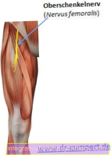
The femoral nerve arises from the lumbosacral plexus, which is composed of the lumbar and sacral plexuses. It arises from the lumbar spine segments L1-L4. The femoral nerve runs down the outer edge of the large lumbar muscle (psoas major muscle) and then runs inside under the inguinal ligament (ilioinguinale ligament) through the muscle box (lacuna musculorum).
Does the Femoral nerve this happens it divides into one in the area of the front thigh motor part (Rr. Muscularis), which supplies the muscles and two sensitive parts (Rr.cutanei anteriores). The sensitive, anterior cutaneous nerve branches (Rr.cutanei anteriores) run through the thigh fascia (Fascia lata) and supply the front and inner surfaces of the skin of the thigh from the groin to the knee. The motor parts (Rr. Muscularis) supply the extensor muscles of the thigh. This includes the pelvic lumbar muscle (M. iliopsoas), where the supplying branches branch off in the pelvis, the four-headed thigh muscle (Quadriceps femoris muscle), the tailor's muscle (M. sartorius) and the comb muscle (M. pectineus). The outer group of the supplying motor nerve group (rami muscularis) supply the lateral part (Vastus lateralis), the thigh extensor (Rectus femoris muscle) and the intermediate part (Vastus intermedius). The middle part (Vastus medialis) is supplied by a nerve branch, which in the middle part of the tailor's muscle (M. sartorius) runs along. In general it can be said that the Femoral nerve supplies all flexors of the hip joint and all extensors of the knee joint.
The fibers that supply the muscles always branch out at different heights into several branches for the different muscle parts. In addition, sensitive fibers branch from these branches to the capsule of the knee joint and the periosteum (Periosteum) of the tibia (Tibia) from. From the nerve branch of the middle part of the four-headed thigh muscle (vastus medialis) branches extend to the vein and artery of the thigh (V. and A. femoralis).
In its course, the femoral nerve is inserted into the Saphenous nerve continues, which provides purely sensitive innervation and, together with the arteria and vena femoralis, through the Adductor canal runs. He runs in front of it Trigonum femoral. Shortly before the end of the canal, it passes through the adductor fascia (Septum intermusculare vastoadductorium). It runs over the inner knee joint gap and supplies the skin on the inside of the lower leg between the knee and Ankle.
Neuroanatomy of the nerve fiber
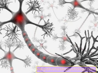
A nerve fiber consists of an axon and a sheath. Basically, a distinction is made between medullary and medullary nerve fibers. The femoral nerve is a medullary nerve. Here the shell consists of the so-called medullary sheath. The myelin sheath consists of myelin, which is formed by the Schwann cells. The myelin sheath is interrupted at regular intervals by constrictions, the Ranvier nodes, which correspond to the expansion of an envelope cell and are called internodes. Saltatory stimulus conduction can take place via these constrictions, in which the stimulus to be transmitted jumps from constriction to constriction and is thus conducted much more quickly than with continuous stimulus transmission. The larger the axon, the thicker its medullary sheath and the longer the individual internodes.
Neuroanatomy of peripheral nerves
The outer (peripheral) nerve fiber is made up of a Endoneural sheath surround. This consists of longitudinal collagen fibrils and the basement membrane. The fibers with their sheaths are in loose connective tissue (Endoneurium) stored. Several nerve fibers are bundled and covered by a further covering structure made of connective tissue (Perineurium) and thus combined to form bundles or fascicles. This layer is reinforced in the area of the joints for mechanical protection. Around the perineurium is another layer of connective tissue, the so-called epineurium. In this course those supplying the nerve Artery, Veins, Lymphatic vessels and annoy, fat cells are also stored. Between the individual connective tissue layers there is a layer of cells (endothelial cells), which ensure that the nerve remains mobile.
The mechanical load capacity results from the content of circular elastic fibers.
Peripheral nerves contain four different types of fiber. For the striated muscles it includes somatomotor fibers, somatosensory fibers for skin sensitivity, visceromotor fibers for the smooth organ muscles and viscerosensitive fibers for the internal organs.


.jpg)