kidney
Synonyms
Renal calyx, renal pole, renal pelvis, renal hilus, migrating kidney, renal cortex, renal medulla, nephron, primary urine, pelvic inflammation
Medical: Reindeer
English: Kidney
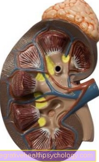
Illustration of the kidney
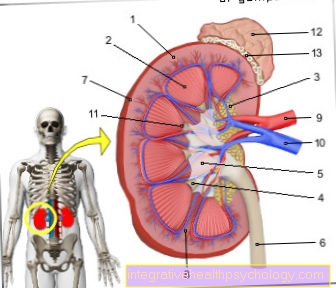
- Renal cortex - Renal cortex
- Renal medulla (formed by the
Kidney pyramids) -
Medulla renalis - Kidney bay (with filling fat) -
Renal sinus - Calyx - Calix renalis
- Renal pelvis - Pelvis renalis
- Ureter - Ureter
- Fiber capsule - Capsula fibrosa
- Kidney column - Columna renalis
- Renal artery - A. renalis
- Renal vein - V. renalis
- Renal papilla
(Tip of the kidney pyramid) -
Renal papilla - Adrenal gland -
Suprarenal gland - Fat capsule - Capsula adiposa
You can find an overview of all Dr-Gumpert images at: medical illustrations
Kidney anatomy
The kidney, of which every human usually has two, is roughly bean-shaped. Each kidney weighs approximately 120-200 g, with the right kidney generally smaller and lighter than the left.
You can find more information about the anatomy of the abdominal cavity here: Abdomen
For orientation of the kidney the doctor describes an upper and a lower kidney pole (upper and lower end of the kidney), an anterior and a posterior surface of the kidney and a medial (i.e. facing the center of the body) and a lateral (outer) edge.
There is an indentation on the medial (inner) edge of the kidney, the so-called renal hilus. This is where blood vessels reach and leave the kidney, and this is also where that is located Renal pelvis, from where the urine passes through the ureter in the bladder got.
The kidney is covered by a tough connective tissue capsule (Capsula fibrosa) overdrawn. Underneath there is a layer of fat, the capsula adiposa, which protects the kidney as it cushions shocks and vibrations.
In the case of severe emaciation (such as a anorexia) this layer of fat can be completely absent, which means that the kidney changes its position due to lack of support (so-called migrating kidney).
The position of the kidneys changes with the position of the body and with breathing: the kidneys are thus lower when standing than when lying down and deeper when breathing in than when exhaling. Due to the space occupation by the Liver (hepar) the right kidney is slightly lower than the left.
Each kidney has its own artery (Renal artery) arising from the Main artery (Aorta) arises and a vein (renal vein) that carries the blood to the inferior vena cava (Vena cava) promoted.
The renal arteries also supply nutrients and oxygen to the adrenal gland, ureters and fat capsules.
Before entering the renal hilum, each divides Renal artery in 2 - 3 branches. Additional kidney vessels are also not uncommon, but are not of any disease value. Nevertheless, knowledge of such unusual blood flow conditions, e.g. B. be important in operations.
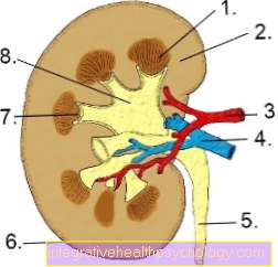
Figure kidney
- Renal medulla
- Renal cortex
- Renal artery
- Renal vein
- Ureter
- Kidney capsule
- Calyx
- Renal pelvis
function

The kidney is divided into:
- Renal cortex and
- Renal medulla.
They are clearly distinguishable in color and structure.
1. Renal medulla (medulla renalis):
The renal medulla consists of approx. 12-15 conical pyramids, the base of which points towards the surface of the kidney, while the tip (papilla) protrudes into the calyx of the renal pelvis. There are several openings in the papilla through which the urine enters the renal pelvis.
2. Renal cortex:
The renal cortex everts itself over the base of the medullary pyramids. In longitudinal sections, the surface appears columnar (so-called Bertini columns). A medullary pyramid with an associated cortical layer forms a kidney lobe that is roughly wedge-shaped.
This is considered the basic unit of the kidney Nephron. It consists of:
- Kidney corpuscles and
- Renal tubules,
which can be divided into different sections of the kidney.
In total, every person has around 2 million nephrons!
1. Kidney corpuscles (glomerulus)
The kidney corpuscle is a tangle of tiny blood vessels (Capillaries), with one inlet and one outlet each (vascular pole). It is surrounded by a capsule (Bowman capsule), which consists of two leaves.
A protein-free filtrate from the blood (the primary urine) is released into the space in between and is passed into a canal system at the urinary pole (opposite the vascular pole).
The walls of the capillaries in the ball have large pores through which the blood can be filtered into the capsule. The passage of protein is prevented by feet cells (podocytes), which cover the pores with their feet like a kind of sieve and prevent the passage of too large particles.
At the vascular pole there is a point of contact with the urinary drainage system, the macula densa. Here the saline concentration of the urine is measured and, depending on the result, the blood flow and thus the filtering performance of the glomerulum is changed.
2. Renal tubules (tubules)
The kidney tubules can be divided into different sections.
- Proximal tubule (main part) with a tortuous and elongated part
The cells lining this channel have a strongly folded surface (brush border). Various enzymes are located here, with channels and pores for the re-absorption of water, sugar (glucose), amino acids, sodium, Potassium, Chloride, phosphate and uric acid are used. The exchange of substances can also take place past the cells through gaps. - Intermediate tubule (transition piece) with descending and ascending part (Henle loop)
The lining cells are flat and have no brush border. Here water is taken up again and the urine is concentrated. This is achieved through an accumulation of table salt in the surrounding tissue, which results in an outflow of water from the tubule. - Distal tubule (middle piece) with stretched and twisted part
It moves upwards into the cortex, where it makes contact with the macula densa at the vascular pole (see above). Here the re-absorption of table salt takes place, which favors the escape of water, and the release of potassium. These processes are under the control of a hormone from the adrenal gland (aldosterone). - Tubule reuniens (connecting tubule)
This is the last section of the nephron. It is tortuous and can accommodate multiple distal tubules.
Several tubules then open into a collecting tube. All twisted tubular sections lie in the cortical labyrinth, all straight in the medulla. - Manifold
In the collecting tube of the kidney, the need-related re-uptake of water and the final concentration of the urine take place under the control of the hormone ADH (antidiuretic Hormon).
The so-called secondary urine (approx. 1.5 - 2 l per day) reaches the renal pelvis from the collecting tubes and then continues through the ureter (Ureter) into the bladder.
Blood supply to the kidney
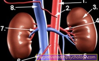
- Abdominal artery (aorta abdominalis)
- Upper intestinal artery (Arteria mesenterica superior)
- kidney
- Renal artery (ateria renalis)
- Ovarian vein / testicular vein (vena ovarica / testicularis)
- Ovarian / testicular artery (arteria ovarica / testicularis)
- Renal vein (vena renalis)
- Lower vena cava (vena cava)
Kidney disease
Kidney cancer
Almost all kidney tumors are so-called renal cell carcinomas. These malignant tumors (malignancies) are relatively insensitive to chemotherapy and can take very different courses. At the Kidney cancer is mostly a tumor of the elderly patient (usually between 60 and 80 years of age).
Further information can be found under our topic: Kidney cancer
Acute kidney failure
The acute kidney failure (ANV) can have various causes, e. B. acute glomerulonephritis, damage to the blood vessels of the kidneys (e.g. vasculitis), toxins and much more. Often it arises after serious injury, surgery, or shock sepsis. In the context of multiple organ failure, the prognosis is particularly poor.
Further information can be found under our topic: acute kidney failure
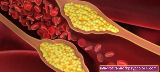
thrombosis
In the constipation an artery (through a thrombosis or embolism) or their branch, e.g. B. by a blood clot, kidney infarction (tissue destruction) occurs in the supply area, which means that the kidney tissue that is no longer supplied with blood dies.