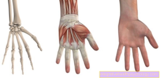
Thigh bone (femur)
Synonyms
Femoral neck, hip joint, knee joint, femoral condyle, trochlea, caput femoris, femoral head, femoral head
English: femur, thigh bone
anatomy
The femur (Femur) is the largest bone in the human body. Like the tibia and fibula, it is a long bone. That means it consists of a hard coat (Compakta) and a soft cavity filled with blood cells (Cancellous bone).
With his femoral head (femoral head = Head femoris) it forms the hip joint with the hip socket of the pelvis. This is a so-called ball joint.
The femoral head is critically supplied with blood. The blood flow takes place in a kind of one-way street from the femoral neck back to the femoral head. Circulatory disorders in this area are therefore more common.
.jpg)
The femoral head is directly connected to the femoral neck. Of the Femoral neck stands at approx. 127 ° to the thigh shaft in an adult person (Femoral shaft).
At the tip of the thigh shaft you can find the on the outside Greater trochanter (large rolling mound) and on the inside the Lesser trochanter (small rolling mound). Both are starting points (Apophyses) for big Muscle groups (Hip flexors and splitters)
The thighbone ends at the knee joint with its two thigh rollers (medial and lateral femoral condyle). These two thigh rolls form with the shin (Tibia) the Knee joint.
Illustration of the femur
_2.jpg)
Thigh bone
Femur, os femoris
- Femoral shaft -
Corpus femoris - Great Rolling Hill -
Greater trochanter - Femoral neck -
Collum femoris - Femoral head (femoral head) -
Head femoris - Headband pit -
Fovea capitis femoris - Small rolling hill -
Lesser trochanter - Inner femoral knot -
Medial epicondyle - Outer femoral knot -
Lateral epicondyle - Articular surface for the kneecap -
Facies patellaris - Bone crest between
the rolling hills -
Crista intertrochanterica - Roughness for the approach of the
gluteus muscle -
Gluteal tuberosity - Rough line -
Linea aspera - Inner joint gnar -
Medial condyle - Intergranular pit -
Intercondylar fossa - External articular knot -
Lateral condyle
You can find an overview of all Dr-Gumpert images at: medical illustrations
_3.jpg)
X-ray image of the knee joint from the front
- Kneecap (patella)
- Fibula
- Thigh bone (femur)
- Shinbone (tibia)
function
The head of the thigh and the socket of the pelvis form the hip joint.
With the condyles of the thigh bone, it forms the knee joint with the shin bone.
As the only bone in the thigh, it transmits all the force from the body (pelvis) to the lower leg (shin / tibia).
Thigh bone diseases
_4.jpg)
Gonarthrosis and coxarthrosis
The most common disease of the thigh bone is osteoarthritis of the knee joint (Gonarthrosis). This is followed by osteoarthritis of the hip joint (Coxarthrosis).
Femoral neck fracture
With increasing age and decreasing bone quality, fracture of the femoral neck (Femoral neck fracture) to. This leads to a break between the femoral neck and femoral head. In most cases, a femoral neck fracture requires surgery.
Artificial Knee Joint / Artificial Hip Joint
A femoral shaft fracture is rare and more likely to occur when great force is used, such as in traffic accidents or a artificial knee joint or artificial hip joint in front.
Fractures above the joint rollers, a so-called one supracondylar femoral fracture also occurs in old age and almost always has to be treated surgically.
_5.jpg)
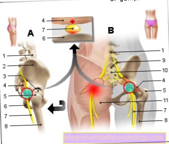
Overview hip
- Beck shovel
- Femoral head
- Acetabulum
- Femoral neck
- Greater trochanter
- Lesser trochanter
Appointment with ?
_6.jpg)
I would be happy to advise you!
Who am I?
My name is dr. Nicolas Gumpert. I am a specialist in orthopedics and the founder of .
Various television programs and print media report regularly about my work. On HR television you can see me every 6 weeks live on "Hallo Hessen".
But now enough is indicated ;-)
In order to be able to treat successfully in orthopedics, a thorough examination, diagnosis and a medical history are required.
In our very economic world in particular, there is too little time to thoroughly grasp the complex diseases of orthopedics and thus initiate targeted treatment.
I don't want to join the ranks of "quick knife pullers".
The aim of any treatment is treatment without surgery.
Which therapy achieves the best results in the long term can only be determined after looking at all of the information (Examination, X-ray, ultrasound, MRI, etc.) be assessed.
You will find me:
- Lumedis - orthopedic surgeons
Kaiserstrasse 14
60311 Frankfurt am Main
You can make an appointment here.
Unfortunately, it is currently only possible to make an appointment with private health insurers. I hope for your understanding!
For more information about myself, see Lumedis - Orthopedists.