Oligodendroglioma
Definition
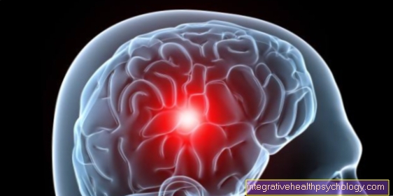
The oligodendroglioma is one of the Brain tumors and is mostly benign. Most often, the oligodendroglioma occurs at the age of 25-40 Years on.
introduction

Oligodendrogliomas are tumors that arise from certain cells of the Brain develop. These cells are called Oligodendrocytes, they surround the Neurons in the brain and serve as a support structure. They are also responsible for the transport of substances and liquids.
Oligodendrogliomas can be divided into four different levels. Ever higher the stage, the more more vicious are they.
One differentiates:
benign oligodendrogliomas, grade I.
low-grade oligodendrogliomas, grade II
anaplastic oligodendrogliomas, grade III
highly malignant oligodendrogliomas, grade IV
The therapy options and survival time change depending on the classification.
Symptoms
The symptoms of various brain tumors are mostly relative similar, since the symptoms are rather through the Location of the tumor is conditional. The oligodendroglioma is mainly in the front brain, this part of the brain is also called Frontal lobe.
If only a small tumor presses the frontal lobe, the symptoms can be very minor. Symptoms in the early stages usually include:
a headache
epileptic seizures
Nausea and vomiting
The headaches are mostly just in the early stages In the morning available. They can also be accompanied by nausea and vomiting. These rather diffuse headaches often get better during the day.
Epileptic seizures are also very common in the early stages, due to their location in the front part of the brain. Since in the front part of the brain there are also the personality of a person sitting, late-stage oligodendrogliomas often lead to Changes of character. These can include:
increased aggressiveness
Drive failure
Poor memory
Neurological failures, like Speech disorders and Paralysis are rare, but possible. This usually only happens when the tumor has reached a certain size and begins to bleed. When that happens, the symptoms occur abruptly up and not slowly creeping in as with a single increase in the size of the tumor.
causes
The cause of the emergence is to this day unexplained. There are many theories, but none of them have been proven. There is evidence that the tendency towards the formation of oligodendrogliomas genetically conditioned can be. There is also a connection with Viruses and Multiple sclerosis discussed.
diagnosis
As with any illness, the diagnosis is first made via the anamnesis, i.e. via a consultation with a doctor. The external history, i.e. the description of the symptoms by a second person, also plays a role in brain tumors. Because often the partner or children notice the changes in personality the quickest. It is also important to give a precise description of the epileptic seizures, as these differ depending on the location and type of tumor.
If the doctor suspects that it may be a brain tumor, an imaging of the head is arranged. A CT (computer tomography) with administration of a contrast medium, an MRI (MRT of the brain) or an X-ray examination of the skull can be done. An EEG (electroencephalography) is also often written, which means that the brain waves are measured.
For diagnosis, the most important are CT and MRI. A CT scan with contrast agent can detect the tumor in over 90% of cases and sometimes also determine the type of tumor.
However, oligodendrogliomas can be seen more precisely in the MRI. The MRI is therefore the method of choice because not only can the type of tumor be better determined, but the location of the tumor can also be displayed very well. This in turn is very important for therapy planning.
If an oligodendroglioma has now been found, a biopsy is usually initiated. The malignancy of the oligodendroglioma can be determined through the biopsy.
Read more on the subject at: Brain biopsy
therapy
Therapy depends on the malignancy, size and location of the oligodendroglioma. The options for treating oligodendroglioma range from Chemo- over radiotherapy to surgery.
As mentioned in the introduction, there are different degrees of this tumor. It ranges from benign to very malignant, with the benign variants much more often occurrence.
If the tumor is now in a location that is easy to reach and is only slightly malignant (grade I or II), an operation may be sufficient. In grade I a complete cure is sometimes possible through an operation, in grade II this is also possible, but less likely. If complete healing is not achieved, radiation therapy or chemotherapy can be prescribed after the operation.
If the tumor is very large or in an unfavorable place, it cannot always be operated on and must be pretreated with radiation or chemotherapy. Radiation or chemotherapy often leads to one reduction of the tumor and it can therefore be operated on afterwards.
If the tumor is more malignant, i.e. grade III or IV, radiation and / or chemotherapy is often used here as well.
Whether an operation, radiotherapy or chemotherapy is preferred and whether these therapy options are combined with one another depends on many factors and must be decided again and again. Aside from the type, location, and size of the tumor, that too play a role Age of the patient who wish of the patient and his Previous illnesses a role.
forecast
The prognosis of an oligodendroglioma depends mainly on the malignancy and the Therapy options dependent. The more aggressive the tumor, the lower the chances of survival. The time of diagnosis also plays a role.
On average, the oligodendroglioma is one slowly, but steadily growing Low malignancy tumor. With good prognostic factors, i.e. with a very good location, small size and low malignancy, the 10 year survival rate at almost 50%. With bad prognostic factors at approx 20%.
The mean survival time is estimated at approx three to five years a. This poor prognosis, despite mostly low levels of aggressiveness, can be explained by the fact that brain tumors have a very large size as they grow pressure exercise on the brain. However, since the brain is dated skull is enclosed, the brain cannot give way to the pressure and is usually severely damaged by the pressure of the tumor.
After successful treatment of an oligodendroglioma, follow-up examinations are very important, as brain tumors often tend to recur, i.e. often recur. Depending on the type of tumor, it may be necessary to take control imaging every 3-12 months. If you are receiving additional or sole chemotherapy, blood tests are also necessary, as chemotherapy can lead to blood formation disorders.


.jpg)

