Optic atrophy
English: optic atrophy
Synonyms
(Optic nerve = optic nerve; atrophy = decrease in the size of the cells, decrease in the number of cells)
Dying of the optic nerve, optic nerve loss
Definition of optic atrophy
Optic atrophy is the loss of nerve cells in the optic nerve. The nerve cells either decrease in size or in number. Both are possible.
Atrophy can have various causes.

Summary
The Optic atrophy describes a loss of nerve cells in the Optic nerve. The nerve cells that the visual impressions of the Retina along the visual path towards brain (Visual cortex), decrease in number or size. This atrophy can have various causes. Some are briefly mentioned: Inflammation of the Optic nerve (optic nerve), increased intracranial pressure, alcohol- or tobacco poisoning.
The symptoms range from unnoticed, small, central ones to large areas and thus restrictive in everyday life Visual field defects.
The trend-setting in the diagnosis is above all Fundoscopy by the ophthalmologist. The treatment of optic atrophy is more difficult because the cause has to be treated. Prophylaxis is just as difficult. The prognosis is also heavily dependent on the various causes and can therefore range from good to bad.
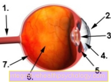
Figure eyeball
- Optic nerve (optic nerve)
- Cornea
- lens
- anterior chamber
- Ciliary muscle
- Vitreous
- Retina
Detecting optic atrophy
What are the symptoms of optic atrophy?
The complaints reported by the patient range from small central defects in the visual field to extensive visual field defects that are very restrictive in everyday life.
The symptoms depend on the cause of the optic nerve damage.
With a special hereditary form (Leber optic atrophy) For example, large central failures occur in the visual field, which cannot be reversed. In optic atrophy caused by tumor pressure, color vision is primarily affected at the beginning of the symptoms, while visual acuity improves again after adequate therapy.
How is optic atrophy diagnosed?
When diagnosing optic atrophy, the ophthalmologist's reflection of the fundus is particularly important. Here the papilla appears (Optic nerve outlet) pale.
Here, too, the diagnosis is easy or difficult to make depending on the cause. The papilla changes impressively in different ways.
Due to the ever better resolution of the MRI, the representation of the optic nerve in the MRI is playing an increasingly important role.
MRT has become more and more established in ophthalmology, especially for assessing the course of the nerves behind the retina / fundus.
Read more on the topic: Optic atrophy MRI
Treating optic atrophy
How is optic atrophy treated?
Treatment of optic atrophy is usually based on the cause. In most cases, however, the therapy is not very promising and there is no improvement in the symptoms.
Treatment is hardly possible, especially in the case of traumatic damage to the optic nerve. Although cortisone tries to reduce the swelling of the nerves, a complete restoration of vision is very often not achieved. If the nerve is compressed by a tumor, a treatment can be found relatively easily by relieving the pressure on the nerve, i.e. removing the tumor.
Can optic atrophy be detected by an MRI?
In order to make the diagnosis of optic atrophy and to be able to better assess its course, it may be necessary to carry out a magnetic resonance tomography (a so-called MRI image). The structures inside the body are made visible with the help of electromagnetic rays.
Due to its molecular composition, an MRI is particularly suitable for displaying soft tissue structures, which is very helpful when assessing the “soft” optic nerve. In this way, the attending physician can assess how far the degeneration has already progressed and whether other space-occupying processes may be the basis of the whole process and also whether the progression of the disease can be slowed down by the therapy or not.
Read more about this at: MRI for optic atrophy
Prevention of optic atrophy
What are the causes of optic atrophy?
Optic atrophy usually occurs as part of or as a result of previous eye diseases.
It is divided into primary and secondary causes:
- primary causes:
This includes all optic atrophies that are not caused by another disease. The optic nerve disc, the point at which the optic nerve emerges from the eye (blind spot), is sharply defined. The following causes are possible:- inherited optic atrophy
- Optic atrophy due to poisoning (tobacco, alcohol, lead)
- secondary causes:
In the case of secondary causes, a disease usually preceded the retina or the optic nerve itself, e.g. Ex. Glaucoma (glaucoma) In these cases, the optic nerve outlet is usually swollen. Often optic atrophy occurs after the following diseases:- Papillitis (inflammation of the optic disc)
- Retrobulbar neuritis (inflammation of the optic nerve behind the eye)
- Congestive papilla (with increased intracranial pressure)
Read more on the topic: Optic atrophy causes
How can optic atrophy be prevented?
Optic atrophy can only be prevented by preventing the cause. Prophylaxis is more or less difficult, depending on the situation. Inherited otic nerve atrophy cannot be prevented, whereas atrophy of the optic nerve from alcohol or tobacco can be avoided.
Course of an optic atrophy
What is the course of optic atrophy?
Optic atrophy is usually a slowly progressing, degenerative deterioration of the optic nerve. The individual nerve cells along the optic nerve gradually degrade, so that the person affected can expect complete blindness at the end of the disease.
In children and adolescents, this process is usually much more accelerated than in patients who only become ill at an advanced age.
According to the current state of science, nerve cells that have died cannot be restored, so that early detection and thus treatment of optic atrophy as early as possible is of great importance. The first symptoms to be noticed by those affected are partial visual field defects and an increasing loss of central visual acuity. Night vision and color perception during the day can also be impaired from time to time.
In imaging procedures, such as ophthalmoscopy, the fundus of the eye shows progressive fading and discoloration of the optic nerve papilla. In order to be able to better assess the damage that has already occurred, further diagnostic procedures such as MRI and VECP are indicated. The earlier the diagnosis of optic atrophy can be made, the earlier a suitable therapy can be initiated and the progression of the disease slowed down or even stopped. If left untreated, however, the disease ultimately leads to complete blindness of the affected eye in almost all cases.
What is the prognosis for optic atrophy?
The prognosis depends on the cause, which is responsible for the optic nerve damage.
If there is a trauma-related cause, experience has shown that the result is bad. In the case of temporary damage to the optic nerve due to tumor pressure, however, the optic nerve recovers surprisingly quickly and well after relief, so that visual acuity is soon restored. In the case of hereditary optic atrophy, the visual loss is irreversible, which means irreparable.
Further questions about optic atrophy
Does optic atrophy also occur in babies?
Optic atrophy in babies and toddlers can have various causes, including a congestive papilla, hydrocephalus, meningioma, retinitis pigmentosa, multiple sclerosis, optic nerve inflammation, traumatic processes and much more.
In Germany, therefore, the eyes of newborns are routinely examined for any incipient pathological changes in order to be able to diagnose and treat accordingly as early as possible. To do this, the doctor uses special eye drops that dilate the baby's pupil and allow him to examine and assess the fundus. He pays special attention to cloudiness and the like. The first signs of discomfort in the child are the inability to fixate objects and people as well as a noticeably strong reaction of the child to light stimuli. If the parents observe this behavior, an early visit to the doctor is advisable.
Can optic atrophy be inherited?
The so-called Leber optic atophy is passed on mitochondrially from generation to generation. This means that the mother is solely responsible for passing on the defective genes, which is why this is also known as "maternal heritability".
Nevertheless, Leber's optic atrophy occurs less often in women themselves. In addition, optic atrophy can also be inherited in the context of other syndromes, such as Behr syndrome I, limb girdle dystrophy 20, motor-sensitive neuropathy VI or Cohen's syndrome. The cause here are malfunctions in the eye on the subcellular level of the tissue.