Bipartite patella
introduction
The patella bipartita is a shape variation of the kneecap that exists from birth, in which it does not consist of one part of the bone, but rather of two separate bone parts due to a disorder in the ossification (lat. bipartite = divided into two parts). This investment variation usually has no disease value, occurs in 2-3% of the population and occurs predominantly in boys or men.

In addition to the two-part kneecap, a three-part (Tripartite patella) or a kneecap made of more than three parts (Patella multipatita) are available. The cartilaginous cover on the side facing the knee joint is not divided into two parts. The bone fragment separated from the rest of the kneecap is most often found in the upper-lateral area, but in some cases there may also be a kneecap that is separated into almost equally sized parts.
Function of the kneecap
The patella is a triangular, flat disk of bone that is in front of the knee joint and plays a key role in its joint surfaces and joint function. It is embedded in the tendon of the greater thigh muscle (Quadriceps femoris muscle) and works as a kind of pulley or placeholder, so that the leverage of the thigh muscle is optimized, the tendon of the muscle is spared and the knee joint is protected.
Read more on the subject at: Kneecap
Figure knee joint
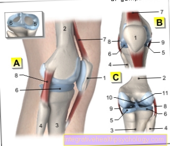
A - Right knee joint from the left
B - Right knee joint from the front
C - Right knee joint from behind
- Kneecap - patella
- Femur - Femur
- Shin - Tibia
- Fibula - Fibula
- Inner meniscus -
Meniscus medialis - Outer meniscus -
Lateral meniscus - Kneecap ligament -
Ligamentum patellae - Outer band -
Ligament collaterale fibulare - Inner band -
Ligament collateral tibial - Posterior cruciate ligament -
Ligament cruciatum posterius - Anterior cruciate ligament -
Ligament cruciatum anterius
You can find an overview of all Dr-Gumpert images at: medical illustrations
Symptoms
A kneecap that is divided into two or more parts does not usually cause any symptoms and is usually only discovered by chance during an X-ray examination of the knee. In a few cases there may be symptoms related to exertion, similar to a disease of the cartilage surface of the kneecap (Chondropathia patellae): First and foremost, it is pain while walking, especially when going downhill, as well as when sitting and squatting for a long time.
Read more on the subject at: Pain in the kneecap
Appointment with a knee specialist?
I would be happy to advise you!
Who am I?
My name is dr. Nicolas Gumpert. I am a specialist in orthopedics and the founder of .
Various television programs and print media report regularly about my work. On HR television you can see me every 6 weeks live on "Hallo Hessen".
But now enough is indicated ;-)
The knee joint is one of the joints with the greatest stress.
Therefore, the treatment of the knee joint (e.g. meniscus tear, cartilage damage, cruciate ligament damage, runner's knee, etc.) requires a lot of experience.
I treat a wide variety of knee diseases in a conservative way.
The aim of any treatment is treatment without surgery.
Which therapy achieves the best results in the long term can only be determined after looking at all of the information (Examination, X-ray, ultrasound, MRI, etc.) be assessed.
You can find me in:
- Lumedis - your orthopedic surgeon
Kaiserstrasse 14
60311 Frankfurt am Main
Directly to the online appointment arrangement
Unfortunately, it is currently only possible to make an appointment with private health insurers. I hope for your understanding!
Further information about myself can be found at Dr. Nicolas Gumpert
diagnosis
Whether the kneecap consists of two or even more bone parts can be determined with the help of an X-ray image. Since the differentiation from a traumatic, old, pseudoarthrotic kneecap fracture does not always have to be clear, performing a bone scintigram can be helpful in unclear cases.
MRI
In most cases, the bipartite patella is discovered incidentally on x-rays. The patella is very clearly visible in the X-ray, so that a patella bipartita can be diagnosed quickly. Often, however, the exact edges of the kneecap cannot be optimally assessed due to overlapping of other bones, which often makes it difficult to distinguish it from a patellar fracture. Therefore, magnetic resonance imaging (MRI) is often performed as an additional measure. This cross-sectional imaging enables a more precise assessment of the tissue and the bony parts of the knee and a demarcation of the bipartite patella from a fracture.
Read more on the subject at: MRI of the knee
root cause
During the embryonic and fetal development in the womb, the kneecap is initially made cartilaginous and then, in the course of growth, remodeled from one point to the bone (ossification).
In some cases, this ossification process can originate from several so-called bone nuclei, with the individual bony structures then merging over time, so that, as a rule, a uniform bone surface is formed. According to earlier assumptions, it was assumed that in the patella bipartita this fusion of the bone nuclei does not occur and that the individual structures do not grow together completely.
In the meantime, however, it is more likely that an ossification of a cartilage system occurs, which is located in an area in which a dent in the bone also occurs at the same time (Emargination).
therapy
Treatment of the patella bipartita is only necessary if it is accompanied by limiting symptoms. If this is not the case, it usually remains untreated, as it does not indicate any disease value.
In the case of stress-related pain, short-term pain relievers and anti-inflammatory agents can be used (e.g. ibuprofen or diclofenac).
In exceptional cases that are refractory to therapy, surgical treatment of the two-part kneecap can also be considered, whereby the unconnected bone fragment is usually removed or - in the case of two-part division with larger parts - the same procedure is used as for the treatment of a fresh patellar fracture: Medium The choice is the use of Kirschner wires in the context of performing a tension band osteosynthesis (the two bone parts are brought together by means of the wires; the tensile force acting on the wires is converted into compressive forces).
surgery
A bipartite patella rarely requires treatment, as it usually does not cause any symptoms. However, if it does lead to pain and if conservative therapy with pain relievers and anti-inflammatory agents has been exhausted, an operation can help.
In most cases, a knee examination is performed first (Arthroscopy) recommended for knee rehabilitation.To remove the painful patella fragment, a small incision is made on the side of the kneecap in the course of the tendon of the vastus medialis muscle. Now the disconnected and painful fragment can be removed. Alternatively, the fragments could also be connected by means of wires as part of the tension belt osteosynthesis, similar to the procedure for a fracture of the patella.
After the operation, the knee is usually fitted with a knee brace for 10-14 days, but full weight bearing is still possible. The knee should also be loaded immediately; if necessary, physiotherapy can help with the healing process. Sport can be resumed after about 4 weeks.
physical therapy
Physiotherapy can be of great help for knee problems. Through targeted exercises and strengthening the muscles of the thigh, symptoms caused by a bipartite patella can be reduced. However, a complete cure cannot be achieved through physiotherapy. If symptoms persist despite muscle strengthening and painkillers, surgical removal of the kneecap fragment is the treatment of choice. But physiotherapy is also of great help in recovery after an operation.