pleurisy
introduction
A pleurisy (pleurisy) is an inflammation of the pleura or pleura (Pleura). The pleura lines the inside of the chest cavity and covers it lung.
Pleurisy is often caused by severe pleurisy Pain noticeable on the affected side, which are usually breath-dependent. Inflammation of the pleura can be a sign of one Underlying disease and severely impair general well-being. In most cases, however, the exact cause is unclear because one invasive diagnostics is not necessary for harmless courses.

Classification of pleurisy
The pleurisy is classified according to various criteria.
It will be a acute onset from one chronic Pleurisy differentiated.
Furthermore, it will be different in severity from one light to a very difficult shape structured.
In addition, pleurisy can be classified according to its cause infectious and one non-infectious form to be grouped.
In the infectious forms, viruses, bacteria or fungi are the cause of the inflammation.
The non-infectious form is caused by pre-existing diseases, such as a Pulmonary embolism, lung infection (pneumonia), Inflammation of the pancreas (Pancreatitis) or due to a Tumor involvement of the pleura (Pleural cancer).
The pleurisy also turns into a dry form (Pleurisy sicca) and a wet form (Exudative pleurisy) differentiated. However, since the dry form also often comes with a smaller one Pleural effusion can go hand in hand, the classification cannot be made so strictly.
causes
The pleurisy is usually a side effect or consequence of an illness or injury.
The most common underlying diseases that can cause pleurisy include pneumonia or an acute infection of the airways in the bronchi. Here, the mostly bacterial pathogens spread from the lungs to the surrounding pleura.
Inflammation of neighboring organs, such as the kidneys, can also be responsible for pleurisy as a microbial source of scatter.
Lung cancer or a pleurus tumor is a potential cause of pleurisy, especially in older people.
Diseases of the cardiovascular system, such as pulmonary embolism, can promote the development of pleurisy. Here, a blood clot washed in from the body's circulation obstructs the pulmonary circulation. The reduced blood flow in this area creates an ideal breeding ground for inflammation.
Tuberculosis, which is a little less common in our latitudes and primarily affects the lungs, can lead to the bacterium sowing into our body's circulation around a year after the initial infection with the pathogen. The consequence is the infestation of other organs, including the risk of pleurisy.
A number of autoimmune diseases, such as systemic lupus, in which the body's own cells are falsely attacked and destroyed by the immune system, can trigger an inflammatory process in the pleura.
Pleurisy is more rarely caused by viruses. A well-known representative that can lead to pleurisy is the so-called Coxsackie virus, the causative agent of Bornholm disease.
In rare cases, "Still's disease", a rheumatic disease, can also be the cause. For the most important information, read the article under: Still's disease - what's behind it?
Gradient forms
The pleurisy can present itself in different forms.
In general, the division into dry or wet pleurisy is based on the fluid surrounding the pleura. The symptomatic symptoms are significantly influenced by this.
Dry pleurisy usually results from pathological changes in the lungs. The leaves of the breast pelts slide past each other with difficulty and thus generate increased frictional resistance, which causes severe breath-dependent pain. As a result, those affected usually adopt a gentle posture and shallow breathing to reduce pain. An accompanying effusion in the area of the pleura is missing. Dry pleurisy can be perceived by the examiner using a stethoscope through a creaking noise caused by the friction.
With wet pleurisy, effusion occurs within the so-called pleural space, which is located between the lungs and pleura. The moist form often arises on the basis of a previously dry inflammation of the pleura. The resulting liquid film, which now separates the two leaves rubbing against each other, reduces the pain. Depending on how much fluid there is in the gap, the damp pleurisy can lead to a feeling of pressure in the chest, difficult breathing and even shortness of breath.
In very rare cases there is a psychological progression of pleurisy. This leads to the development of the typical symptoms of pleurisy with no apparent cause. The influence of mental processes such as sadness, stress or depressive moods can be reflected in the form of these physical illnesses.
You may also be interested in this topic: Pleural mesothelioma
infection
There are two different forms of pleurisy (pleurisy).
There is a infectious and a not infectious Form of pleurisy. The non-infectious form is not infectious and can arise as part of other inflammatory processes in the body. So it is possible that they are under one of the Inflammation of the pancreas (Pancreatitis) develops. It is also possible that non-infectious pleurisy is a concomitant phenomenon Pulmonary embolism or one Pleura tumor is. Even patients who attend rheumatism sufferers can develop non-infectious pleurisy due to their underlying rheumatic disease.
The infectious form pleurisy is contagious depending on the pathogen. The pathogens can be bacteria, Viruses or Mushrooms act. The bacterial pathogens are usually the same as one lung infection (Pneumonia), so this form is just as contagious as pneumonia. Also the causative agent of tuberculosis can trigger pleurisy. These bacteria are only very contagious when, for example, through to cough can be easily disseminated. There is also an increased risk of direct contact persons infected with viruses Risk of contagionwhich, however, through a regular and thorough disinfection can be greatly reduced. In the case of an infection with fungi, the risk of infection is usually only with people in contact with one defective immune system elevated.
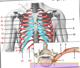
I-XII ribs 1-12 -
Costa I-XII
(I-VII) True ribs -
Costae verae
(VIII-X) false ribs -
Costae spuriae
(XI-XII) rudimentary ribs -
Cost. fluctuantes
- Rib head - Caput costae
- Rib neck - Collun costae
- Rib humps -
Tuberculum costae - Rib body - Corpus costae
- Rib head joint -
Articulatio capitis costae - Sternum - sternum
- Vertebral bodies -
Corpus vertebrae - Costal cartilage -
Cartilago costalis - Rib-sternum joint -
(Sternocostal joint) - Rib-vertebral body joint -
(= Point 5.)
You can find an overview of all Dr-Gumpert images at: medical illustrations
Symptoms
The leading symptom of pleurisy is breath-related pain. This can be localized in the entire chest cavity and is particularly pronounced when inhaling. Patients often describe this as a palpitation of the heart when inhaling. If the pleurisy is accompanied by a pronounced accumulation of fluid in the pleural space (pleural effusion), in some cases there is no pain at all, since the pleural leaves do not rub against each other. The effusion can also make breathing difficult with shortness of breath.
In the dry form with no or only a slight effusion, the pleural leaves rub directly against each other and thus lead to severe pain and typical auscultation findings: When inhaling, the examiner hears a so-called “leather creak” with a stethoscope. In addition to these symptoms, symptoms of pleurisy can also be fever, fatigue, shortness of breath, throat irritation and a reduced general condition.
Duration of symptoms
The duration of symptoms in pleurisy varies widely. Depending on which underlying disease triggered the inflammation and when appropriate therapy was initiated.
Pleurisy due to a flu-like infection usually heals without consequences after a few weeks with appropriate therapy.
Pleurisy due to an underlying malignant disease can be much more difficult to treat.
If the inflammation persists for a long time, scars can appear between the pleura and the pleura, which permanently restricts breathing.
Duration of pain
Pain is most common with dry pleurisy. When you breathe, the pleural layers rub against each other and cause stabbing and burning pain. Those affected breathe short and shallow.
If the inflammation and irritation of the pleura persists for a long time, fluid can form, which collects in the space between the pleura and the lungs, causing a so-called pleural effusion and moist pleurisy. From this point on, those affected no longer have pain.
With appropriate treatment and administration of painkillers, the symptoms usually also disappear after a few days. Adequate pain therapy is essential for healing. It is important that those affected breathe deeply in order to adequately ventilate the lungs. Due to the pain, this is often not possible with dry pleurisy, so pain relievers should definitely be taken.
Diagnosis of pleurisy
The basic diagnosis consists in the History taking and description of typical complaints, such as pain or pressure in the chest, relieving postures or shortness of breath.
As part of the physical Examination with a stethoscope the doctor stops changing Breath sounds of the patient. This is typical of dry pleurisy Pleural rubbing (Leather creaking) to be heard, with the damp form becoming a Decrease in the sound of breathing can come. in the Ultrasonic Pleural effusion and irregular lung contours may be seen as signs of inflammation. Also can fever, Laboratory values and especially the Inflammation values again CRP value Provide evidence of pleurisy.
If the exact cause is not known, a X-ray image performed by the lungs to rule out pneumonia.
Recently, the representation of the lungs in the MRI has made great progress, so that in special cases a MRI of the lungs should be thought.
To a Pulmonary embolism can exclude a Vascular presentation in the legs be helpful and if the inflammation values persist and the fever is high, become one Blood culture and a Pleural puncture carried out.
Here the pleural effusion is punctured and the material is exposed bacteria, Viruses, fungi and tumor cells are examined. This is often the way to find the cause.
This invasive measure is only necessary if the symptoms do not improve. An uncomplicated one pleurisy does not have to be punctured.
The X-ray examination
The roentgen belongs next to that Ultrasonic one of the diagnostic methods for pleurisy. With pleurisy usually forms between lung and diaphragm something liquid (Pleural effusion). In an x-ray taken in Hard blasting technique a radiologist can assess how much fluid is present. With this recording technique, the bones are poorly shown and the soft tissues are shown well.
Duration
The duration of the pleurisy (Pleurisy) is highly dependent on what triggers the disease. At a bacterial cause the disease can with a favorable course within a few days heal. If the course is less favorable, the disease can drag on for several weeks. However, if the cause is not an infectious event, the disease can extend over a longer period of time. Is the cause for example rheumatic genesis, the underlying disease must first be treated so that the pleurisy can improve. As in pleurisy liquid can collect between the lungs and diaphragm, a so-called Pleural effusion, it can help symptomatically through a drainage to empty and so relieve the surrounding tissue. Especially for patients who are affected by the inflammation shortness of breath suffer, this can be helpful. A particularly lengthy form of pleurisy treatment is chosen when the pathogen is tuberculosis bacteria. Depending on whether it is an open form of the tuberculosis, i.e. contagious tuberculosis or not, the patients are then treated in the hospital for a while.
For more information, read our topic: Duration of pleurisy.
Duration of dry pleurisy
Dry pleurisy usually heals faster than a wet one. Since the underlying disease is often an acute event, such as you have a flu-like infection or lung disease. With appropriate therapy, the inflammation disappears with treatment of the underlying disease.
In otherwise healthy people, dry pleurisy usually heals without consequences after a few weeks.
With damp pleurisy, the underlying disease is often more difficult or impossible to treat, e.g. Autoimmune diseases, lung cancer, heart failure. Even with appropriate therapy, new pleural effusions can form here again and again.
therapy
Treatment of pleurisy is primarily symptomatic.
Pain medication and cough suppressant juices can relieve symptoms.
If the cause is bacterial inflammation, antibiotics are given.
Mycotics can be given in the event of fungal attack. However, this is rarely the case. If there are cancer cells in the pleural effusion, the pleura can be treated with chemotherapy. In this case, however, the basic therapy is treating the primary tumor.
In some cases, for therapeutic reasons, it may be necessary to puncture the effusion. This is always useful if the effusion is so large that it displaces the lungs on the affected side and breathing no longer works properly. By draining the effusion, the lungs have space again in the chest and can expand.
Most pleural effusions, however, are relatively small and self-resorb over time, so there is no need for a puncture. Nevertheless, the actual cause must be treated with medication, otherwise the effusion will reappear quickly.
If the pleural effusion is purulent, it can also make sense to temporarily place a drain in the pleural space. In this way, the purulent effusion is continuously drained and the inflammation can subside. Such a therapeutic measure only makes sense in combination with a systemic antibiotic.
In any case, patients should take care of themselves and not engage in strenuous activities or sports as long as the pleurisy is not completely healed.
During sleep, it is advisable to lie the patient on the healthy side so that the sick side is better ventilated. However, since patients often have pain and difficulty breathing, many patients tend to lie on the sick side in order to get better air.
In the event of breathlessness, oxygen can also be administered through a nasal tube.
Duration of treatment
The duration of treatment depends on the underlying disease. A flu-like infection with accompanying pleurisy usually only needs to be treated for a few weeks. A malignant disease such as Lung cancer requires lengthy radio therapy, chemotherapy and possibly surgical measures. Here the development of a relapse (flare-up of the inflammation) is common.
Home remedies for pleurisy
Home remedies can help if you have mild pleurisy as they can relieve symptoms. One option here is to make warm chest wraps.
Another possibility is hay flower therapy. The compresses with the moist, warm hay flowers are applied to the affected area for almost three quarters of an hour. It is important that the hay flowers come from a pharmacy to ensure that there are no impurities or harmful substances on the plants. The home remedies work purely symptomatically, but not causally, so if the symptoms do not improve despite the home remedies, you should definitely consult a doctor to avoid spreading the disease. Since many patients take gentle breathing because of the pain, it is important that they start breathing training at home and breathe deeply despite the pain so that the less well-ventilated sections of the lungs are ventilated and so that pneumonia cannot develop.
More information can be found here: Chest wrap
Exercise for pleurisy
Basically, patients who suffer from pleurisy initially no sport should do before the cause of the disease is known. If it is a pleurisy from the area of the infectious forms, it makes more sense to begin with save. The body needs energy to fight an infection successfully, so the body should be treated during one acute infection do not put heavy loads on it. The most sensible thing is to listen to your own body awareness and not to put yourself under full strain again immediately after the illness, but rather to slowly increase the training to the level it was before the illness.
In the case of the non-infectious form of pleurisy, it is important to practice sport to the extent that it is beneficial to your health. So a light endurance training instead of strength training. Here too, of course, you have to learn to pay attention to the signs of your body and to react. In case of doubt, one should not do one during the acute illness very strenuous sport and slowly start again after the illness.
danger
The risk of pleurisy is spreading it. It can be too Adhesions of the pleura, so that the agility the lungs are restricted. Another danger is that a Pleural effusion can become so large that the Lung volume comes and then becomes massive as a result Shortness of breath. Therefore, if you suspect pleurisy, you should consult a doctor as soon as possible.
forecast
As a rule, pleurisy without complications heals completely without any problems.
If the pleurisy is caused by another disease, the prognosis depends on the healing of the disease.
In some cases, pleurisy heals as a scar, so that smaller strands of scarring in the pleural space can lead to adhesions and pain.
These can usually easily be separated surgically.
The prognosis of untreated pleurisy due to tuberculosis is very rare today, but can lead to death, as can an untreated purulent inflammation of the pleura.
Summary
Inflammation of the pleurisy (pleurisy) is an inflammation of the pleural leaf, which can be caused by viruses, bacteria or fungi, but also by other diseases such as pneumonia or tumor disease.
Inflammation of the pleura is associated with severe pain, especially breath-dependent, and can massively worsen the general condition.
The diagnosis is made with the help of the medical history, a physical examination and auscultation, as well as laboratory results. An ultrasound examination can be used to visualize an effusion.
If there are indications of non-inflammatory causes of the disease, the entire body must be examined.
The therapy is mainly symptom-oriented and is carried out with pain medication and cough suppressants.
Antibitoics are given if the bacterial inflammation is severe.
In some cases it is also necessary to perform a pleural puncture or insert a pleural drainage.
The prognosis is favorable in most cases, but if left untreated, severe bacterial inflammation or tuberculous pleurisy can lead to death.


.jpg)