skull
definition
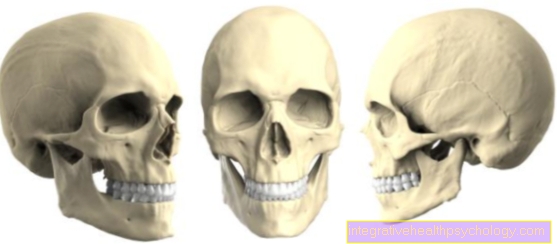
As a skull (Latin: Cranium) one describes the bony part of the head, so to speak the head skeleton.
Bony structure
The human skull consists of many bonewhich, however, through the Bone sutures (Sutures) are firmly fused together.
These seams belong to the spurious joints. In the course of life these sutures gradually ossify, one then speaks of Synostoses.
Immediately after birth, however, some of the bone sutures are not fully formed. Here are Bone gaps, the so-called Fontanelles, which is why the head of newborns feels soft in certain places because there is no bone here. During the first years of life they close Fontanelles usually in a certain way. However, if the bones grow together incorrectly in the further course, characteristic changes in the shape of the head occur, for example for
- Scaphocephalos ("Boat-shaped") or to
- Trigonocephalos (triangular).
Figure skull
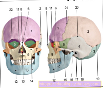
- Frontal bone -Frontal bone
- Parietal bone - Parietal bone
- Occiput - Occipital bone
- Temporal bone - Temporal bone
- Sphenoid bone - Sphenoid bone
- Ethmoid - Ethmoid bone
- Zygomatic bone - Os zygomaticum
- Nasal bone - Nasal bone
- Upper jaw - Maxilla
- Lower jaw - Mandible
- Tearbone -Lacrimal bone
- Chin hole - Mental foramen
- Ploughshare - Vomer
- Under eye cavity hole -
Infraorbital foramen - Zygomatic arch -
Arcus zygomaticus - Temporomandibular joint -
Articulatio temporomandibularis - External ear canal -
Meatus acousticus externus - Mastoid process
(Part of the temporal bone) -
Mastoid process - Lambda seam -
Sutura lambdoidea - Cuticle seam -
Sutura squamosa - Crown seam - Coronal suture
- Orbital upper edge -
Margo supraorbitalis
You can find an overview of all Dr-Gumpert images at: medical illustrations
Classification
For anatomical purposes, the skull is divided into two parts:
- the Brain skull (Neurocranium) and
- the Facial skull (Viscerocranium).
The brain skull consists of 8 bones:
- the unpaired Occiput (Occipital bone),
- the paired Parietal bone (Parietal bone),
- the paired Temporal bone (Temporal bone),
- the unpaired Sphenoid bone (Sphenoid bone),
- part of the Frontal bone (Frontal bone) and the
- unpaired Ethmoid bone (Ethmoid bone).
In addition, the roof of the skull (skullcap, calvaria) can be distinguished from the base of the skull in the neurocranium.
About two Head joints there is direct contact between the Brain skull and the Spine. The Spinal cord exits through an opening in the base of the skull and runs in the spinal canal in the spine to the coccyx. The bones of the brain skull house ours brain and represent an important protection of the brain against external influences. In addition, the brain is not directly on the bone, but is again in a liquid (dem Brain water or Liquor) embedded so that shocks or the like can be better absorbed.
Facial skull
Of the Facial skull is formed from the following bones:
- The proportions of the Frontal bonewho have an involvement in the eye socket,
- the paired Zygomatic bone (Os zygomaticum),
- the originally paired upper jaw (Maxilla),
- the paired Intermaxillary bone (Os incisivum),
- the unpaired Lower jaw (Mandible),
- the paired Nasal bone (Nasal bone),
- the paired Nasal concha bone (Os conchale),
- the paired Tear bone (Lacrimal bone),
- the paired Palatine bone (Palatine bone),
- the unpaired Ploughshare (Vomer) and
- the unpaired Ethmoid bone (Ethmoid bone).
The bones of the facial skull form the basis of our face and thus determine to a large extent how we look.
While the ratio of brain to facial skull is still around 8: 1 in newborns, it is only around 2: 1 in adults.
Skull base
The Skull base refers to part of the brain skull (Neurocranium). In contrast to the Facial skull (Viscerocranium) the skull directly surrounds that brain and thus fulfills a certain protective function. The base of the skull is now that lower portion this brain skull, it is made up of several bony parts. They participate in the construction Sphenoid bone (Sphenoid bone), the Temporal bone (Temporal bone), the Frontal bone (Frontal bone), the Ethmoid bone (Ethmoid bone) and the Occiput (Occipital bone).
The base of the skull is allowed, however not as a flat structure imagine because of the walnut-like shape of the brain, it can be divided into three pits. It is furthest towards the face anterior fossa (Anterior cranial fossa), in the area of the back of the head there is the posterior fossa (Posterior cranial fossa) and exactly between the anterior and posterior fossa you will find one middle fossa (Fossa cranii media).
Each of these pits has characteristic holes (Foramina) on. These holes serve as passages for various annoy, Arteries and Veins, besides, can every cranial fossa assign a section of brain.
In the anterior fossa (Anterior cranial fossa) is mainly the front part of the brain (Frontal lobes) and the, important for the smell, Olfactory nerve. It is formed by that Frontal bone (Frontal bone), Parts of the Ethmoid (Ethmoid bone) and sections of the Sphenoid bone (Sphenoid bone).
The middle fossa (Fossa cranii media) is mainly bounded by the sphenoid and temporal bone, it mainly includes the lateral portion of the brain (Temporal lobe) and the Pituitary gland. Most of the penetration points are located in it and thus the central fossa also has the most connections to other cavities of the bony skull.
The main connections are:
- Optic Canal (between the base of the skull and the eye socket), this is where the Optic nerve (Optic nerve) and that artery that contains the eye socket and the eye self-sufficient (Ophthalmic artery).
- Superior orbital fissure (between the base of the skull and the eye socket), through which especially the Eye muscle nerves (Occulomotor nerve, Trochlear nerve and Abducens nerve) and the sensitive Nerve of the upper half of the face (Ophthalmic nerve) pull.
- Foramen rotundum (between the base of the skull and the palatine fossa) through which the Maxillary nerve (Maxillary nerve) occurs.
- Foramen oval (leads pathways from the base of the skull out of the skull) with the Mandibular nerve (Mandibular nerve).
The posterior part of the base of the skull, formed by the posterior fossa (Posterior cranial fossa), is limited by Parts of the temporal bone and the occiput. There are more in this section of the skull base small depressions to recognize. This is what happens in these hollows Cerebellum and the venous outflow routes (Sine) to lie.
Within the posterior fossa are mainly Connections to the ear (on the Porus acousticus internus) and to the spinal canal (about the Foramen magnum). On the Porus acousticus internus get both the Hearing and equilibrium nerves to the inner ear. The Foramen magnum lies completely in the occiput and represents the most important connection between the brain and the spinal canal because both the elongated brain stem and the Meninges, and the pathways supplying the spinal cord pass through this opening in the base of the skull.
On the basis of the anatomical conditions just explained, it can be understood why a Categorized skull base fracture as life-threatening is.
By Violence, mostly in the course of Traffic accidents, it comes to fracture (fracture) the anterior, middle and, in rare cases, the posterior fossa. Common symptoms are severe a headache, Vomit, the exit of blood and Cerebral fluid (Liquor) from the nose or ears and impaired consciousness.
Skull bones
As Skull bones are all bones of the human skeleton above the Cervical spine designated. They can be roughly divided into those surrounding the brain Brain skull bones and shaping the face and jaw Facial skull bones. The brain skull consists of the Occiput (Occipital bone), the two Parietal bones (Parietal bone) such as Temporal bones (Temporal bone), as well as the Sphenoid bone (Sphenoid bone) and the Frontal bone (Frontal bone). At birth, these are not yet completely fused together, but this happens during the first two years of life.
With the exception of the articulated Lower jaw (Mandible) the bones of the facial skull in adults have grown together. Aside from the lower jaw bone, there are only those Ploughshare (Vomer) and the Ethmoid bone (Ethmoid bone) Bones of the facial skull that are placed in the middle and therefore only occur once per person. The Nasal bones (Nasal bone), lower turbinates (Concha nasalis inferior), the Palatine bones (Palatine bone), the Cheekbones (Os zygomatic), the Tearbones (Lacrimal bone), as well as the Maxillary bone (Maxilla) appear twice, symmetrically placed on the left and right.
If this Hyoid bone (Os hyoideum) and the Auditory ossicles Malleus (hammer), Incus (anvil) and Stapes (stirrup) also pay to the bones of the skull is controversial. In their entirety, the cranial bones are among the most variable in shape in the human skeleton. A large number of skull shapes can be distinguished on the basis of various measuring points.
Injuries

The skull is obviously a vital structure of our body, which is why injuries to the skull should always be taken seriously.
Common injuries are, for example
- Traumatic brain injury and the
- Base of skull fracture.
In the case of a traumatic brain injury, an external influence causes an injury to the skull with brain involvement. This trauma can be visible from the outside, then one speaks of an open head trauma. Here are
- the scalp,
- the skull and
- possibly the hard meninges (Dura mater) to see.
Brain tissue may leak out of the head laceration. In the same way, a traumatic brain injury can be closed, one also says covered.
This is by no means less dangerous!
Despite the lack of external evidence, it can be massive
- Cerebral hemorrhage,
- Crushing or
- Swellings appear, which in the worst case can lead to unconsciousness.
The symptoms do not have to appear immediately after the trauma, which is why it always makes sense to monitor a patient who has suffered a traumatic brain injury in the hospital in order to rule out complications.
The skull base fracture (Base of skull fracture) is also caused by violence against the head, often from a traffic accident. Most often the fracture gap is either in the area of the nose or the ear. Depending on this, brain water often leaks either from the nose or the ear. In patients with a fracture of the base of the skull, one can often see hemorrhage around one or both eyes (corresponding to monocle or glasses hematoma), as escaping blood can easily collect in the soft tissue behind the eyes. There are both mild and severe courses that correspond to the picture of traumatic brain injury.
traumatic brain injury
Will be part of a injury (mostly by accident) both the Skull bones, as well as that brain affected, so the expert speaks of a Traumatic brain injury (SHT).
Depending on whether the external force was affected Meninges (Dura mater) is broken or not, it is either a more severe open SHT or a covered trauma.
A further distinction is made as to whether the impact of violence directly the brain injured (direct damage), or whether about through Bleeding or Swelling As a result of the injury, the brain is in distress.
Depending on an assessment of the state of consciousness of the TBI patient based on the so-called Glasgow Coma Scale (GCS), for which a maximum of 15 points can be achieved, the clinician assesses the severity of the TBI. A GCS value of 13-15 points corresponds to a SHT grade 1 (concussion) no permanent brain damage is to be expected. With 8-12 points in GCS it is one Brain contusion (SHT grade 2). Prolonged unconsciousness and more pronounced symptoms than with a concussion are typical. Below 8 points on the GCS scale indicate a so-called Brain contusion (SHT grade 3). In order to be able to counter the severe injuries to the brain that this entails, at least partially in a healing manner, the person concerned often remains Weeks in unconsciousness. Complete restoration of all brain functions is possible, but very unlikely.
Skull MRI / MRI of the head
The Magnetic resonance imaging of the skull, also Magnetic resonance imaging called, is a radiation-free imaging Procedure, which is mainly used in medicine to assess soft tissue structures. In comparison to the method of the CT, but with X-rays works and, above all, better depicts bony structures, the MRI is louder, more expensive and takes much longer (e.g. 10 to 30 minutes for a skull MRI). In emergencies, an MRI image of the head is therefore usually not taken.
For other questions about (any) Diseases of the skull or the inside of the skull, especially the soft tissue, where there is no such time constraint, an MRI examination is often the imaging method of choice.
This is next to one increased expressiveness Above all, this can be explained by the renunciation of X-rays or other ionizing (and thus potentially carcinogenic) rays. However, since the examination requires the patient to lie still for many minutes in a very noisy, narrow tube, some people find the MRI of the head uncomfortable.
The magnetic resonance tomograph works with rapidly changing, strong Magnetic fieldsthat align primarily hydrogen nuclei in one direction in the body, such as a large comb. If these then jump back into their original alignment, this creates a small electromagnetic pulse that is measured. Depending on how and how much hydrogen is bound in a tissue, this so-called resonance signal decreases in strength and time delay to the "Comb“Differently off and on Image contrast arises. Depending on the weighting of the signals, either high fat, or but water-rich Fabric in the picture bright.
With an additional venous administration of Contrast media, in this case Gadolinium, the information content of an MRT image series can be increased further. Especially when looking for tumorous tissue or Foci of inflammation a so-called additional contrast medium sequence is of inestimable diagnostic value.
Common indications for a Skull MRI are therefore the suspicion of a tumor-like event (such as a lump in the brain, or else Resettlements (Metastases) an original tumor located elsewhere) and the suspicion of one inflammatory process (e.g. as part of a Multiple sclerosis).