Visual pathway
introduction
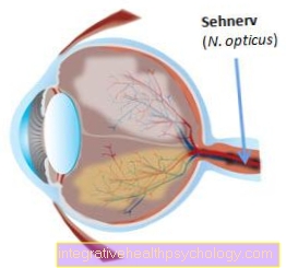
The visual pathway is part of the brain because all of its components originally come from it, including the optic nerve. The visual pathway begins at the retina (retina), the ganglion cells of which are the starting point, and ends in the visual cortex in the cerebrum. Their complex structure enables us to see.
Anatomy of the visual pathway
The structure of the human visual pathway is very complex. It begins at the posterior pole of each eye and ends in the cerebral cortex of the cerebrum.
The first nerve cells that belong to the visual pathway can be found in the retina. The ganglion cells of the retina unite to form Optic nerve (Optic nerve) and emerge from the eye socket (orbit). The optic nerve consists of two different parts of fiber bundles. If you look at the retina, it can be divided into a lateral (outer) and medial or nasal (inner, towards the nose) part.
If you look at the beginning of the visual path from above, the following results: In the right eye, the lateral part of the retina is on the right and the nasal part on the left, while in the left eye it is exactly the other way around. Understanding this fact is essential for understanding the further course of the visual path.
First, the fiber bundles of the nerve cells of the retina of the respective eye attach to each other, sometimes cross, only to reunite a little later in another combination.
The branching point is called Optic chiasm. Only the fibers that represent the nasal parts of the retina cross here. So after the intersection run in the so-called Optic tract on each side the fibers of the corresponding sides of the retina.
The right optic tract now guides the fibers of the right halves of the retina, the left tract those of the left halves. In other words: the uncrossed fibers of the right eye and the crossed fibers of the left eye now unite in the right optic tract. These retinal sections correspond to the left halves of the field of view. The uncrossed fibers of the left eye and the crossed fibers of the right eye unite in the left Optic tractwhich corresponds to the right halves of the field of vision.
Course of the visual path
The visual path extends from the retina of the eyes to various areas of the brain. The most distant area of the brain lies on the back wall of the skull and thus on the head on the opposite side of the eyes.
The beginning of the visual pathway is represented by the sensory cells of the retina, the rods and cones. The first visible part of the visual pathway is formed by the emerging optic nerve, the optic nerve. This nerve is first visible at the back of both eyes.
From there, the optic nerves run back through the middle of the eye socket and form the so-called optic chiasm, the junction of the visual pathways, in front of the brain stem. This is where the nasal fibers of the optic nerve cross. The nerve fibers are referred to as the optic tract.
An optic tract opens into the diencephalon on each side. From here fibers lead to the primary and secondary visual cortex.
What is the visual path crossing?
The visual path crossing occurs at the point where the visual pathways of both eyes meet. This lies between the eye sockets and the brain stem. At the junction of the visual pathway, the middle, nasal nerve fibers cross over to the opposite side. The outer, temporal nerve fibers remain on their side and do not cross.
After crossing the visual pathway, each visual pathway contains a portion of nasal and a portion of temporal nerve fibers. As a result, the stimuli from the right half of the field of vision are processed in the left brain and the stimuli from the left half of the field of vision are processed in the right brain.
Function of the visual pathway
The visual pathway is used to transmit visual impressions and signals from the eye to the brain. The transport of this information, which is converted into electrical signals, is necessary in order to perceive the visual impressions. If what we see was not transmitted into the cerebrum, we would not be able to perceive what we see.
The visual pathway is also coupled with the sense of balance and the adjusting reflexes. If the impression of the eye deviates from that of the organ of equilibrium, this is balanced out by the adjusting reflexes. On a ship that fluctuates in the sea, both the eyes and the organ of equilibrium / vestibular organ perceive the fluctuations and activate the corresponding muscles so that we continue to stand firmly.
If you're more interested in this topic, then check out our next article below: How does vision work?
Insertion of the field of view
The retinal sections reflect the fields of view in an opposing arrangement. The right part of the visual field of each eye is recorded on the left side of the retina. The left halves of the visual fields are accordingly mapped onto the right parts of the retina.
The right and left tracts are switched in the midbrain. From here the so-called visual radiation moves into the cerebral cortex. It ends in the occipital lobe on the inside of the two halves of the brain (hemispheres) in the visual center.
You can find out more about examining the visual field under: Examining the visual field
What is the consequence of a failure of the visual pathway?
Injury to the visual path has roughly always formulated a complete or an incomplete loss of the field of vision as a consequence. If the secondary visual cortex is affected, stimulus processing is impaired. Depending on the location of the injury, the loss of the field of vision takes different forms.
If the injury is in front of the optic chiasm, a complete eye will fail. If the injury is in the optic chiasm, the visual field will be on the same side of both eyes. If there is an injury to the visual path after the optic chiasm, the loss of the field of vision can vary greatly.
The damage to the visual pathway can be divided into three sections: prechiasmal, chiasmal and retrochiasmal diseases.
In prechiasmal disease, the optic nerve is mainly affected. A one-sided visual disorder, such as blindness or visual field loss, occurs on the side of the respective lesion.
The chiasmal disease is located at the level of the union of both optic nerves, the so-called chiasma opticum.
This usually occurs when a tumor in the pituitary gland (pituitary adenoma) presses on this structure.
The patient then typically has a so-called bitemporal hemianopia, also known as the blinker phenomenon, since the external field of vision fails on both sides.
Retrochiasmal diseases describe damage that affects the sections after the union of both optic nerves. A typical clinical picture is the homonymous hemianopia: Here the fields of vision on the same side of both eyes are affected.
What is Chiasma Syndrome?
Chiasm syndrome has three components and occurs when the junction of the visual pathway along the midline is damaged. This leads to a conduction disturbance in the middle parts of the retina and the field of vision of the outer sides of both eyes is no longer perceived. In addition, visual acuity is often reduced.
This can occur on one or both sides. Since the nerve cells of the optic nerve can no longer be fully used, the injured nerve cells continue to be lost.
You can read more detailed information on this topic under: Chiasm Syndrome