Tendons
Tendons are used to transmit tension between muscles and bones. They represent the fibrous end piece with which the muscle attaches to its bone. The attachment points are usually as bony protrusions (Apophyses) visible on the bone. These have to be particularly resistant, as they absorb the force transmitted by the muscle via the tendon.
In addition to the normal insertion and origin tendons, there are also intermediate tendons that connect two bellies of a muscle, as well as flat tendon plates (Aponeuroses), such as. on the sole of the foot and the palm of the hand.
The ligaments that connect the moving parts of the skeleton with each other must be separated from the tendons. In contrast to tendons, they are stretched between two bones and serve to stabilize the skeletal system. Tendons consist of tight connective tissue, namely collagen fibers and a few elastic fibers. The whole tendon is in turn surrounded by a layer of loose connective tissue, which ensures its anchoring and at the same time its mobility. The interior of the tendon is divided into individual fiber bundles by fine layers of connective tissue, which, among other things, serve as nerve-vascular roads. Overall, however, tendons contain only a few vessels and nerves, which explains why they are so poorly able to regenerate.
There are two types of tendons, the Compression and tension tendons.
- Tension tendons are subject to tension and consist of tight connective tissuethat is aligned parallel to the corresponding pulling direction.
- Pressure tendons are subjected to pressure and pull in contrast to the tensile tendons around the bone. The bone serves as an abutment. On the side adjacent to the bone, these tendons consist of fiber cartilage, which is not supplied with blood.
To better gliding ability some tendons, especially those that run directly on the bone, are from one Tendon sheath (Synovial vagina) surround. This is similar in structure to a joint and contains a small amount of liquid that the Increased gliding ability of the tendon. This can also reduce the friction between tendons and bones. Tendon sheaths are mainly located around the Tendons of the hand and foot muscles. With heavy use (e.g. always the same hand movement when writing), the Inflamed tendon sheaths (Tendovaginitis).
Tendons are generally very tear-resistant. They transmit with all movements great forces on the bones. But tendons also have one Spring action. With passive stretching, they store part of the energy and release it again during the movement to be performed. Movement sequences can be clearly understood designed more efficiently because the muscles do not have to apply all the strength on their own.
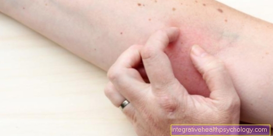
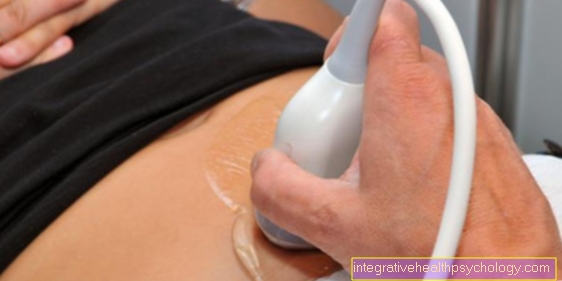
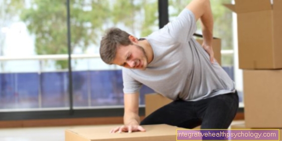
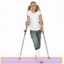
Due to the high load, the tendon tissue is subject often signs of wear and tear (degenerative changes). Injuries to the tendons are often caused by Incorrect loads, twisting or shear forces in the context of Sports injuries. In the case of minor injuries, it is usually sufficient to immobilize the affected region, in the case of larger injuries or even a tear of the tendon operated on become. It usually takes time for the skin to heal completely 4-6 weekswhereupon the tendon but spared a few more months should be.
The main tendons
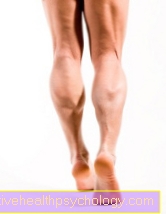
Achilles tendon
The Achilles tendon (lat. Tendo calcaneus) is the strongest tendon in the human body. she holds Loads of up to 800kg was standing. Their length is between 20 and 25 cm and it connects the three-headed calf muscle (Triceps surae muscle) with the heel. Above all, it enables the foot to flex towards the sole of the foot (Plantar flexion), as well as lifting the inner edge of the foot (Supination). These movements are elementary in walking and running. This is also important in this context Achilles tendon reflexwhich can be checked by moderately striking the tendon with a reflex hammer. If the reflex can be triggered, the foot should move Bend towards the sole of your foot. This can be used to check whether the Spinal cord in the amount of Sacral region (Segments S1-S2) works properly.
In athletes, the Achilles tendon is more often affected by injuries. It should be a complete Demolition of the tendon come (Achilles tendon rupture), this is mostly expressed in one loud, whip-like bang. With young people there is usually a operational approach indicated in the elderly is also partially Treated conservatively (Immobilization, Painkiller). There is also excessive stress on the Achilles tendon Obesity and orthopedic foot deformities (e.g. Buckle foot).
Historical background: The Achilles tendon takes its name from the Greek mythology receive. Achilles was there a mortal son of a human father and the sea nymph Thetis. She dived her son into a river, the Styx to make it invulnerable. She held him by the heel, which prevented the water from getting wet from the river. This so-called Achilles heel remained his only vulnerable point.
Quadriceps tendon
The Quadriceps tendon connects the great Thigh extensor muscles (Quadriceps femoris muscle) with the Kneecap (patella). Accordingly, it serves the Power transmission from the thigh to the knee area, as well as the fixation of the kneecap, which is embedded in the quadriceps tendon in its natural position is held. The quadriceps tendon is then continued in the lower knee area by the patellar tendon, which ultimately transfers the force to the lower leg. At sporting activitiesthat involve running, jumping, and frequent abrupt braking can result in a Inflammation of the quadriceps tendon come (Tendonitis).
The complete Quadriceps tendon avulsion can also through strong, abrupt braking movements to be triggered. As a rule, however, the tendon has already been damaged. The injury is usually treated surgically and ends in a long-term immobilization in a cast and subsequently intensive training.
Please also read our article on this Tendinitis in the leg
Patellar tendon
The Patellar tendon (lat. Ligamentum patellae) pulls as Continuation of the hamstring tendon (Quadriceps femoris muscle) of the Lower edge of the kneecap (patella) to their point of attachment on the anterior tibia. The tendon is about 7cm long and 5-6mm thick. Your main job is Power transmission from the thigh to the lower legso that a powerful extension of the leg (Extension) is possible. Besides, she gives that Knee joint stability and fixes the kneecap in its proper position. The patellar tendon is found in various sports often affected by injuries. A complete demolition is rare. Rather, it comes from overloading the tendon tiny cracks (Microcracks) in the tissue. This is mostly expressed through Tenderness in the area of the lower kneecap, as well as through Pain when stretching affected leg. This clinical picture is also called Patellar tip syndrome designated. Treatment is usually carried out by reducing the load and giving anti-inflammatory drugs.
The patellar tendon can also become Checking the functionality of the spinal cord can be used, namely by triggering the Patellar tendon reflex. This is what the tendon does with bent knee tapped with a reflex hammer. If the reflex can be triggered, the Lower leg forward fast. The spinal cord segments checked are in the Lumbar region (Segments L2-L4).
Biceps tendon
Of the Biceps muscle at the upper arm (Biceps brachii muscle) has some special features. He has one big and a small head, each with its own tendon in the area of the shoulder arise from. The joint insertion of both heads lies in the large biceps tendon in the crook of the elbow, where they are attached to one Roughening of the spoke (radius) starts. This distal biceps tendon is about 22mm long and 7mm thick. Your job is above all that Power transmission from the upper arm to the forearm and with it the Flexion of the elbow joint (Flexion). In addition, it serves the Outward rotation of the forearm (Supination). To check the associated Biceps tendon reflex after placing the examiner's thumb on the tendon, this region can also be tapped with a reflex hammer. If the reflex can be triggered, there will be a Contraction of the biceps muscle, where the Elbow joint is bent. The biceps tendon reflex characterizes the Functionality of the spinal cord segments C5-C6 at the bottom Neck area.
In addition to the distal biceps tendon, there are also the both tendons of origin of the heads of the biceps muscle. The tendon of the large head, the proximal (near the trunk) long tendon, plays a role. It primarily serves the Inward rotation and splaying of the shoulder joint. The tendon runs over the head of the humerus in its own tendon sheath under the roof of the shoulder and is especially common in old people often affected by degenerative changes. It happens with increasing age Signs of wear and limescalewhich is why this tendon causes pain or can even tear. The long biceps tendon is also often damaged by excessive strain in sports that place heavy strain on the shoulder (e.g. baseball).
The tendon of the small head of the biceps muscle is less frequent from injuries affected. Its main purpose is to bring the upper arm closer to the body (Adduction). If the long biceps tendon fails, for example through Rupture, the short biceps tendon can do most of the work, leaving it to only one Power loss of about 15% comes. This is also the reason why the long proximal biceps tendon is ruptured in many cases not surgically reversed but the tendon is simply in its incorrect position fixed on the upper arm becomes.
Triceps tendon
The Triceps tendon connects the Triceps muscle on the upper arm with the elbow. The tendon radiates with some fibers into the joint capsule of the elbow joint and serves that purpose stabilization. There is a under the triceps tendon Bursa, of the prevent excessive friction between the tendon and bone should.
The main job of the triceps tendon is Power transmission between the upper arm and ulna (Ulna), mediating the extension of the elbow joint (Extension). This function can through Triggering the triceps tendon reflex be checked. This is what the tendon does just above the elbow (Olecranon) tapped with the reflex hammer. The reflex that can be triggered is expressed by a Extension in the elbow joint. The Spinal cord segments C7 and C8 in the lower neck area.
Because the triceps very superficial runs on the arm, it can in an accident slightly injured become. A rupture of the triceps tendon is very rare and even causes the rarest tendon injury in orthopedics. Usually such a tear occurs only if the tendon is already damaged or falling on the arm, the tendon then mostly bony, i.e. along with a piece of the elbow, rips off. The rupture is treated usually operational.
Rotator cuff
The Rotator cuff is a Muscle tendon platethat of the tendons of the four shoulder rotators is formed and surrounds the shoulder joint. The muscles involved are Muscle supra- and infraspinature, Subscapularis muscle and Muculus teres minor. These muscles take care of that Inward and outward rotation of the shoulder joint and stabilize this in its position by the formed tendon plate. This is important because the shoulder joint is only very slightly secured by ligaments and therefore on a increased muscular fixation is instructed.
Injuries to the shoulder can lead to Tendon ruptures come in the area of the rotator cuff (Rotator cuff rupture). Because the tendons in the shoulder area exposed to heavy loads signs of wear and tear are also common. The therapy of injuries depends on their extent. At a complete demolition the rotator cuff must operated in any case become.