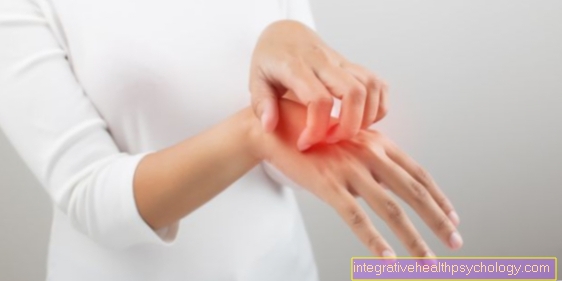
Tendon tear
synonym
Tendon rupture
introduction
The tendon is called the connective tissue parts our muscles. Tendons are there to unite the respective muscles Origin or approach to bones or other muscles and the Power transfer from the muscle to allow on the skeleton. Structurally speaking, a tendon is made of tight connective tissue and emerges seamlessly from the ends of the muscles. The muscles can therefore access various structures via the tendons, but mostly at bone be fixed. For the connection of two or more muscles without bony involvement one speaks of Intermediate tendons. The muscle belly of the respective muscle is divided into 2 parts by the intermediate tendon. In general, the tendons can also be in Tensile and sliding tendons subdivide.
This differentiation he follows according to the course depending on the direction of action of the muscle:
A Tension tendon is only stressed by tensile load, because it has the same course as the muscle and therefore the direction of action is the same.
A Sliding tendon is on the other hand loaded not only by tension but also by pressure, because it does not have the course of the direction of action of its muscle.
Like all structures of the musculoskeletal system can also Tendons be harmed. It plays besides inflammatory or degenerative changes of the Tendon tear a major role. This can be a tendon completely or only partially tear. Due to various properties, tendons are generally very prone to rupture. For one, are tendons not very elastic and on the other hand, they have one insufficient ability to regeneratebecause they are poorly innervated and supplied with blood. The nutrition takes place via a special tissue fluid that is located around the tendons. In most cases, tendons attach to bony structures, so that their involvement in a tendon tear is not atypical. One then speaks of one Avulsion fractureif in addition to the tendon tear also the bone is broken at the point of attachment of the tendon.
causes
Even if tendons are not very elastic, tendons do not tear when subjected to extreme stress. First of all, you can Tendons stretched / overstretched become. If, however, a certain tolerance limit of the tear strength is exceeded, the rupture occurs. Depending on the severity, the tendon will only tear partially or completely, possibly including a bone tear.
The causes of a tendon tear are variable. Because the tendons are closely related to their respective muscles, are especially certain movements or forces cause of a tendon lesion. Sudden loads with extreme pressure and tensile loads can lead to a tendon rupture. In general, strong external acts of violence, such as a blow to the tendon area, a tendon tear.
A tendon is also particularly susceptible if it is under tension at the time of the force or force being exerted, or if the load is inclined. Certain factors or conditions can Increase risk of tendon rupture. This also includes, for example, the inevitable Aging process, the one Loss of elasticity of the tendons all over the body.
Another risk factor is the fact that already damaged tendons are more susceptible and a tendon tear is more likely. Such prejudicial reasons can inflammatory processes or Overload be. Overloading plays an important role in sports. Often small lesions remain on the tendon, e.g. Small partial tears, undetected, and due to more and more stress without regeneration time, the risk of a complete tendon tear increases more and more.
Apart from this creeping injury process, it can of course also increase in sport acute tendon ruptures come. Torn tendons caused by trauma are generally the most common. But not to be forgotten either degenerative and inflammatory Processes that are considered a risk factor for a tendon tear.
Appointment with ?

I would be happy to advise you!
Who am I?
My name is dr. Nicolas Gumpert. I am a specialist in orthopedics and the founder of .
Various television programs and print media report regularly about my work. On HR television you can see me every 6 weeks live on "Hallo Hessen".
But now enough is indicated ;-)
In order to be able to treat successfully in orthopedics, a thorough examination, diagnosis and a medical history are required.
In our very economic world in particular, there is too little time to thoroughly grasp the complex diseases of orthopedics and thus initiate targeted treatment.
I don't want to join the ranks of "quick knife pullers".
The aim of any treatment is treatment without surgery.
Which therapy achieves the best results in the long term can only be determined after looking at all of the information (Examination, X-ray, ultrasound, MRI, etc.) be assessed.
You will find me:
- Lumedis - orthopedic surgeons
Kaiserstrasse 14
60311 Frankfurt am Main
You can make an appointment here.
Unfortunately, it is currently only possible to make an appointment with private health insurers. I hope for your understanding!
For more information about myself, see Lumedis - Orthopedists.
Special locations
A tendon tear can be found in different parts of our body. Typical places of manifestation are described below. At the upper extremity the tendons of the shoulder region, forearm and hand are particularly affected.
Shoulder region and forearms
A tendon tear of the supraspinatus tendon often occurs on the shoulder. The supraspinatus tendon is part of the eponymous important muscle of the shoulder joint, the M. supraspinatus. Together with three other muscles, it forms the rotator cuff, which is essential for the stability of the shoulder joint, as it is a muscle-secured joint. In general, the rotator cuff is particularly affected by a tendon tear, but the long biceps tendon can also tear in the shoulder area. The supraspinatus tendon, however, is very susceptible to lesions due to its course. This is because it passes between the bony roof of the shoulder and the head of the humerus and has its point of attachment to a certain bone process (Greater tuberosity) of the humerus. This tightness can lead to a bottleneck syndrome, the so-called impingement syndrome, which can be associated with severe pain. Further damage from overstressing or signs of wear and tear can weaken the supraspinatus tendon so much that it ultimately tears. It is therefore important to treat warning signs such as impingement syndrome to prevent a tendon rupture.
Read more about this on our website Torn tendon in the shoulder
Further down the arm, the tendons of the forearm muscles are popular places of manifestation of a tendon tear. Since we have many muscles on the forearm that function both as extensors and flexors and are divided into superficial and deep muscle groups, it is too complex to explain each individual muscle now. They all have the above causal factors in common that lead to a tendon rupture. Sports such as weightlifting, gymnastics, wrestling, javelin throw and shot put are also particularly dangerous.
Supraspinatus tendon tear
The supraspinatus muscle belongs to the shoulder muscles and is part of the so-called rotator cuff, a group of muscles that move and stabilize the shoulder joint together. The supraspinatus muscle is mainly responsible for external rotation and abduction (the lateral removal of the upper arm from the body) of the upper arm. The tendon of the supraspinatus runs in the shoulder joint in the narrowness between the shoulder roof bone and the humerus head. As a result, the tendon is exposed to strong mechanical stress, which can lead to rupture due to chronic incorrect stress, the aging process or an accident.
Often times the symptoms develop gradually over months or years. In addition to pain when the arm is loaded, patients often complain of shooting nocturnal pain. An experienced orthopedic surgeon can suspect a supraspinatus tendon tear with the help of a physical examination. However, the gold standard for the final diagnosis is an MRI scan.
The supraspinatus tendon rupture can be treated both conservatively (including physiotherapy, pain therapy) and surgically. An optimal solution should be found for each patient individually.
Also read:
- Rotator cuff tear
Biceps tendon tear
The biceps muscle is located on the upper arm and is responsible for flexing the elbow joint. A biceps tendon tear can affect either the long, upper, or lower biceps tendon. A rupture of the long biceps tendon (also called proximal rupture) is the most common form of biceps tendon rupture and usually occurs as a result of banal accidents with chronic previous damage.
A rupture of the long biceps tendon is usually relatively painless, the strength and function of the arm is only slightly restricted. The biceps muscle shows up as a bulge above the elbow joint, so it practically slides down.
The lower biceps tendon tears mainly in acute injuries, often as part of sports injuries. It is accompanied by acute, stabbing pain. It is not uncommon for a large hematoma (bruise) to form. The flexion in the elbow joint is limited, the outward rotation of the hand is usually no longer possible.
In addition to the physical examination, an ultrasound examination can also be used diagnostically. If trauma is the cause of the biceps tendon tear, an X-ray can be useful to rule out fractures. A rupture of the long biceps tendon is usually treated conservatively with pain therapy and rest. A tear in the short biceps tendon is always treated surgically.
Torn tendon at the crook of the elbow
Tendons can also tear in the crook of the elbow. This is the lower tendon of the biceps muscle, which starts from the upper arm and attaches to the spoke of the forearm via the crook of the elbow.
The tear of the lower biceps tendon is rather rare and occurs mainly in acute injuries, especially among athletes. The rupture is very painful and a bruise often forms. The flexion in the elbow joint is mostly weakened, the outward rotation of the hand is usually completely eliminated. A reliable diagnosis is possible using MRI or ultrasound. Therapeutically, the tendon is surgically fixed again.
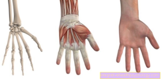
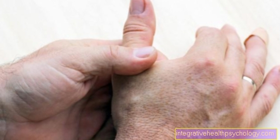
Localization on the hand
However, the individual localizations on the hand, i.e. on the respective fingers or thumb, are now important.
On the fingers are like during sports activities Volley, hand and basketball especially the tendons of the extensor muscles at risk. These can be done at the 3 joint regions Finger joint, Central joint or Base joint but also minor injuries tear due to strong pressure and tensile loads.The individual tendon tears have different therapeutic indications; so a tendon tear of the finger end joints, also "Hammer finger" designated, rather conservative using a finger splint treated.
However, if the tendon ruptures in the Area of the central joint, must be operated on, as the risk of permanent functional impairment is too high and the tendons cannot heal adequately through immobilization alone. The misalignment resulting from a tendon tear of the finger extensor in the middle joint is called "Buttonhole deformity" designated. The result is a characteristic picture, since the fingers can be stretched in the end joint, but can only be bent in the middle joint due to the torn tendon.
In a few cases, lesions in the area of the metacarpal joint relate to Extensor hood. This is a holding system for all tendons that pull over the base joint into the fingers. In the case of strong external forces, it is not so much a single tendon in the base joint that tears, but rather the extensor hood tears. Surgical care is essential here. Characteristic you can when closing the fist a "snapping sound“Perceive if the extensor hood is torn.
The long extensor tendon of the thumb often ruptures M. extensor pollicis longusso that the extension in the end joint is no longer possible.
Read more about finger tendon tears here.
Localization of the lower extremities
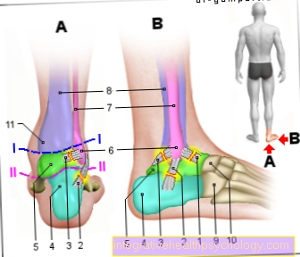
There are also special locations on the lower extremity where the occurrence of a tendon tear is typical. The ruptures manifest themselves at the transition from the lower leg to the foot.
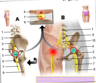
First is the Achilles tendon (Tendo calcaneus) to call. Due to its ability to withstand a tensile load of more than 1 t, it is the strongest tendon in our body. It is particularly at risk when doing sports. Sports such as skiing, tennis and jogging are predisposed. If the Achilles tendon tears, there is a loud bang, which can be compared to a whip.
Furthermore, the tendon of the M. tibialis posterior, part of the deep calf muscles, can be affected by a tendon tear in the foot region. The muscle pulls from the back of the lower leg along the inner ankle towards the bottom of the foot and is responsible for flexion and supination in the foot. However, a total tear is rather rare; most of the time there are maximally fine longitudinal tears in the tendons. Rather, degenerative processes and overload also play a role as an acute trauma. In the course of a tendon lesion, tendinitis comes first, which is caused by overstrain, overstretching and degeneration. The consequences are swelling, pain and ultimately small longitudinal tears.
The malalignment of the foot “buckled foot” also increases the risk of tendinitis. With regard to the tendon of the tibialis posterior muscle, the clinical picture "Tibialis posterior dysfunction“, A degenerative change in the tendon at its point of attachment with a consequence of the loss of function is more frequent and more significant than a tendon tear.
Finally, the peroneal tendon of the peroneus longus muscle, the long fibula muscle, should be mentioned. In contrast to the tibialis posterior tendon, this tendon runs along the outer malleolus. What the tendons have in common, however, is that the total tear is a less common injury here too. An important disease is the so-called "Peroneal Tendon Split Syndrome". Only a fine longitudinal tear of the tendon of the peroneus brevis muscle occurs in the area of the outer malleolus; the long peroneal tendon is not torn, but “drills” into the longitudinal tear from behind. So one can say that there are several places of manifestation for a tendon tear distributed on the body with different degrees of severity and significance.
Read about this too: Tendinitis of the tibialis posterior tendon
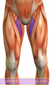
Torn tendon on the thigh
Tendons can also tear in the thigh. This mainly affects the tendon that connects the large thigh muscle (quadriceps) with the kneecap. Typically, this tendon tears when there is an impending fall when climbing stairs due to the jerky tension of the quadriceps muscle.
A rupture manifests itself with pain and swelling in the knee area. In addition, the extension in the knee joint is severely restricted. The torn tendon in the thigh is diagnosed through an ultrasound scan or an MRI. Surgical suturing of the tendon helps therapeutically.
Symptoms
The symptoms of a tendon rupture are usually very typical. Relatively at the same time as the rupture event uses sudden and stabbing pain in the corresponding tendon region a. Since the pain is very severe, a tendon tear is noticed very quickly compared to other musculoskeletal injuries. The only exception is the partial tendon rupture: In this case, it is a small one Microlesionwhich is often only temporarily painful and does not bring the classic symptoms with it. After the pain has subsided, however, the tendon tear has by no means healed, so that there is often further stress with an increased risk of tendon rupture. This creeping change in the end mostly ends with a Total tear the tendon.
Classic symptoms for the tendon rupture are next to that severe pain, the swelling and hematoma formation (bruise). In addition, the Move through the pain but above all through the Loss of function restricted, since the tendons are there for that, the power of the muscles on the skeleton transferred to. If a tendon is completely torn, the muscle lacks the decisive attachment point, so that despite contraction due to shortening, no movement can result. If you inspect the affected area of the tendon tear, you can also see a bruise and swelling in some cases Retraction or dent detect. This is due to the fact that the tendon is no longer attached to its actual attachment point or its continuity is interrupted by the rupture. As a result, the muscle cord with the tendon as a "branch" does not represent a continuous and uniform structure, but rather forms an interruption in the form of a dent or indentation at the rupture site.
All of the above Symptoms vary of course in yours intensity and Duration depending on location and size the torn tendon. The movement restriction is also more or less registered depending on the importance of the affected muscle in the movement. As an example, consider comparing the Achilles tendon and a finger extensor tendon. A tear in the Achilles tendon makes it almost impossible to take the strain while walking. The rupture of a finger extensor restricts the respective movement to a certain extent, but the significance or the extent of the movement restriction is not comparable. Regarding the size of the ruptured tendon, the occurrence of the tendon rupture can even be audible: Tear them Achilles tendon As the strongest tendon in the body, there is a loud bang, as already mentioned, which can be compared to a whip.
Diagnosis
To recognize a tendon rupture or to diagnose it correctly is already a detailed anamnesis interview crucial. Those affected can provide important information about a tendon rupture through a detailed description of the possible course of the accident. It is the task of the attending physician to inquire about the occurrence of typical symptoms and to investigate them.
The Pain intensity can be inquired about Painfulness can through Palpation the affected tendon can be checked. At this point the doctor can next to the Tenderness a possible Dent or Confiscation determine.
If a patient reports a pop-like noise followed by severe pain and swelling of the particular tendon region, a tendon rupture is very likely. To secure the diagnosis of tendon rupture, it is helpful in a Investigation to check functionality to the resulting Assess restricted mobility to be able to. There are certain function tests for this, depending on which tendon of the body is affected. In doing so, attention was paid to movement restrictions, instabilities and abnormal mobility.
In addition to the anamnesis and the examination are also imaging procedures relevant to diagnosing a tendon rupture. These include the X-ray, the ultrasound examination and, if necessary, the MRI (Magnetic resonance imaging). The use of the 3 imaging methods varies depending on the location or severity. If there is a large tear in the tendon, which can already be diagnosed with relative certainty by the inspection, the ultrasound examination helps to visualize the sinewy structure and the tear. If you suspect that, in addition to the tendon tear, the bone is also broken, i.e. an avulsion fracture is present, an X-ray image in 2 planes can provide information about this, as the bony structures can be clearly shown in an X-ray image. Finally, an MRI can be prescribed if necessary. With the help of MRI, even the smallest structural changes can be detected in different cutting planes, so that this imaging method is superior to X-rays and ultrasound in complicated cases. However, x-rays and ultrasound examinations are initially standard.
Therapy and prophylaxis
A tendon tear can be treated both conservatively and surgically.
The conservative treatment also includes immediate measures according to the PECH rule (Break, ice, compression, elevation). If the person concerned feels sudden severe pain with a previous popping noise and subsequent swelling of the corresponding region, the current load should be paused immediately in order not to cause any further damage or to provoke a total tendon tear if, for example, there is only a partial tear at the current point. Then it is important to cool with ice, to compress the region with the help of tight bandages and to elevate the area of the tendon. The aspects of the PECH rule are ideal for first aid and are beneficial for the further healing process of a tendon tear. Depending on the severity, a tendon tear can heal again by consistently resting. When the torn ends are close enough together, small tears or partial tendon tears can grow back together.
A tendon tear can always be treated with medication. So-called anti-inflammatory drugs can be given against inflammation. Common painkillers are effective against pain. Since there may not be much movement in the first few days after the operation, many patients are prescribed thrombosis prophylaxis. It is used to keep the blood fluid enough so that the lack of exercise prevents blood clots from forming, which could loosen and trigger a pulmonary embolism in the lungs.
In addition to drug prophylaxis, preventive measures can be taken so that the tendon does not tear in the first place, or the risk of this is kept lower. Before doing any sport, you should, for example, integrate a sufficient warm-up program including stretching into the training to prepare the tendons for the strain.
Another aspect relates to the nutrition and vitality of the tendons. The tissue fluid mentioned at the beginning ensures the nutrition of the tendons, but this is rather slow. This sluggish diet can be accelerated and improved by increasing activity in the form of regular exercise. Good circulation strengthens the tendon and prevents it from becoming brittle and less elastic than it already is. Regular exercise and stretching can reduce the risk of tendon rupture.
In order to optimize the healing process, it makes sense in some cases to use certain support systems. If the Achilles tendon is torn, it is good to wear a raised shoe, as this relieves the tendon and has a better chance of healing. If the tendon tears in the hand, wearing a splint can help the healing process.
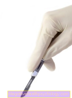
Tear tendon surgery
In most cases, however, this conservative type of therapy is not sufficient, so that an operation must be performed. It makes sense to operate, especially for athletes, as the tendon will be exposed to heavy loads again after recovery and the risk of a renewed rupture is too high with purely conservative therapy.
The aim of the surgical treatment is to sew the torn tendon parts back together and fix them. A tendon must either be anchored again intraoperatively at its starting point, usually on the bone, or sewn together along its course if the continuity of the tendon has been interrupted. There are very specific suturing techniques or stable ones in order to adequately reattach the tendon Titanium anchor which can be buried in the bone. If there is an avulsion fracture, the broken bone must be treated in addition to the torn tendon. The screwing of the splintered bone portion to the bone is particularly suitable here. The entire procedure can be minimally invasive, depending on the location.
If tendons in a joint region are affected, the operation can be performed arthroscopically. This means that only 2-3 small incisions are made and a camera and a trocar with the necessary instruments are inserted. Such an operation method can be used, for example, in the case of a tendon tear of the shoulder, the supraspinatus tendon. After each tendon rupture operation, the affected area must be immobilized for 4-6 weeks. Then it is important to slowly bring the tendon back to the load and not to put extreme stress on it all of a sudden.