Tendon sheath
definition

The Latin technical term for the tendon sheath is "vagina tendinis". A tendon sheath is a tubular structure that surrounds a tendon like a guide channel, for example to guide it around a protruding bone. A tendon sheath protects the tendon from mechanical injuries.
construction
A tendon sheath consists of two layers. The outer layer will Stratum fibrosum called the inner one Synovial stratum. The stratum fibrosum consists of strong collagen-containing connective tissue and is with the structures of the environment firmly grown together. These adhesions attach the tendon sheath and the tendon it contains to bones and ligaments.
The stratum synoviale is made up of an outer, wall-related sheet and an inner, visceral sheet. The outer sheet is also made of connective tissue and lies between the stratum fibrosum and the inner leaf, which is directly connected to the tendon. There is one between the stratum fibrosum and the stratum synoviale Lubricantthat one too Synovial fluid or Synovial fluid is called. The stratum fibrosum and the stratum synoviale are connected by the so-called mesotendin, a strand of connective tissue. Run in this mesotendineum Blood vessels and annoy to supply the tendon and the tendon sheath.
Occurrence
Tendon sheaths always occur when tendons are in Near the joints or must be routed around protruding bones and tethers, which occurs only on the long tendons of the muscles of the arms, hands, legs and feet.
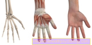
Tendon sheaths of the hand
The hand can perform many different movements and has a high level of fine motor skills. This wide range of motion is made possible by many different muscles and joints. The tendon sheaths of the palm are from the Carpal band (Ligamentum carpi transversum) covered. The superficial and deep finger flexors (Musculus flexor digitorum superficialis or profundus) have one common tendon sheaththat runs under this tape. This tendon sheath only partially envelops the tendons of these two muscles for the 2nd, 3rd and 4th fingers and does not enter the finger itself, the tendon for the little finger however, it is completely enveloped by this tendon sheath and only ends at the terminal phalanx of the 5th finger. The 2nd, 3rd and 4th fingers have a new tendon sheath to protect the tendon in the area of the Finger joints.
The long Thumb flexors-Muscle (M. flexor pollicis longus) has its own tendon sheath, which also runs under the carpal ligament and only begins on the Thumb distal phalanx ends. Also the flexor carpi radialis muscle, which has a flexion in the wrist has its own tendon sheath under this ligament. The tendon sheaths of the second to fifth fingers are attached to the knuckles by ring-shaped bands. In addition, the ligament apparatus is stabilized by ligaments running in the shape of a cross at the joints.
In 70% of the cases, the anatomical position of the tendon sheaths described here is found on the palm of the hand with a long tendon sheath for the thumb and little finger and interrupted tendon sheaths for the three remaining fingers. Anatomical variations so come at 30% of the population before and have no disease value.
At the back of Hand run the tendons of the Finger extensor and the tendon sheaths surrounding them in so-called Tendon fansunder a strap called the extensor retinaculum musculorum. in the first The long thumb spreader (M. abductor pollicis longus) and the short thumb extensor (M. extensor pollicis brevis) are located in the tendon compartment. The first tendon compartment is subdivided again in many people.
in the second The long and short radial hand extensors (M. extensor carpi radialis longus and brevis) are found in tendons.
The third The tendon compartment houses the long thumb extensor (M. extensor pollicis longus) and im fourth The common extensor muscle of the 2nd-5th Fingers and an additional extensor muscle for the index finger (M. extensor digitorum or indicis).
The little finger also has an additional extensor muscle (M. extensor digiti minimi), whose tendon is in the fifth Tendon fold runs.
in the sixth The tendon of the ulnar hand extensor (M. extensor carpi ulnaris) runs through the tendon. In the area of the fingers are found on the extensor side no More tendon sheaths, because the tendons of the muscles on the back of the hand merge into one Connective tissue plate, the Dorsal aponeurosis, skip. This dorsal aponeurosis begins in the area of the metatarsophalangeal joints and ends at the distal phalanx of the respective finger.
Tendon sheaths of the foot
The muscle bellies of the long muscles of the foot are located on the Lower leg, the tendons must therefore around the Indoor or. Outer ankle be redirected. To protect against mechanical damage caused by friction on the bone, the tendons are therefore in the area of the Ankle joints provided with tendon sheaths. The tendons of the long and short fibula muscles (M. fibularis longus or brevis) run behind the outer malleolus and are held by a tether, the Retinaculum musculorum fibularium secured so that they cannot shift or twist. The tendons of the extensor muscles are located on the back of the foot.
Under the Retinaculum musculorum extensorum run the tendons of the long toe extensor and the long big toe extensor (M. extensor digitorum or hallucis longus) as well as the tendon of the anterior tibial muscle (M. tibialis anterior).
The tendon of the posterior tibial muscle (M. tibialis posterior) runs along with the tendons of the long toe flexor and the long big toe flexor (M. flexor digitorum or hallucis longus) behind the medial malleolus. The Retinaculum musculorum flexorum protects the flexor muscles from lifting. The long toe flexors also have tendon sheaths on the sole of the foot to protect against mechanical damage.
More tendon sheaths
In addition to the many tendon sheaths on the hands and feet, there are other tendon sheaths on the elbow and the long one Biceps tendon in the shoulder area.
Tendinitis
A Tendinitis (Tendovaginitis) can be caused by either one infection or by a Overload the tendon arise. The tendon sheath swells and is very painful because there are many sensitive nerves in a tendon sheath. A special picture of tendonitis is that V phlegmon: In the palm of the hand the tendons of the 2nd-5th Fingers in a common tendon sheath, which is in the immediate vicinity of the tendon sheath of the long thumb flexor. The tendon sheaths of the little finger and thumb envelop the entire tendon up to the distal phalanx of the respective finger. Through the tight proximity can Infections In this area, spread it slightly from the little finger over the ball of the hand to the thumb, thus imitating the shape of a "V", which is what gave this special form of tendinitis its name.
Non-infectious Tendonitis is caused by a Over- or Improper loading the affected tendon. There is a more fibrinous Effusion, so a swelling with fibrous parts. These fibrous fibrin coverings are deposited on the inner sheet of the stratum synoviale. Due to the now rough (instead of actually mirror-smooth) surface, the gliding ability of the tendon is reduced and when moving the fingers, rubbing can be felt in the palm of the hand, for example.
People who work a lot on computers, musicians, craftsmen or athletes who play tennis or golf are particularly affected. A special form of non-infectious tendinitis is Tendovaginitis stenosa: Aging processes of the connective tissue lead to thickening of the tendon sheaths in the area of the metatarsophalangeal joints, the thumb is most frequently affected. These thickenings reduce the tendon's ability to slide and hinder mobility. If this handicap is overcome by increased effort, the finger suddenly jumps forward, which leads to the concept of "flicking finger“Led.