Esophageal cancer
All information given here is only of a general nature, tumor therapy always belongs in the hands of an experienced oncologist!
Synonyms
Esophageal Carcinoma, Esophageal Tumor, Esophageal Tumor, Esophagus-Ca, Beret Carcinoma
definition
The esophageal cancer (esophagus = esophagus) is a malignant, uncontrollably fast growing tumor that originates from the cells of the esophageal mucosa.
In 80-90% of cases there is a connection between long-term consumption of high-proof alcohol (alcohol abuse) and the consumption of cigarettes. The esophageal carcinoma can also develop from a beret esophagus, which is a consequence of reflux disease (chronic heartburn). The tumor causes symptoms late, when it is already well advanced. Due to the late diagnosis, this type of cancer has a very poor prognosis for patients.

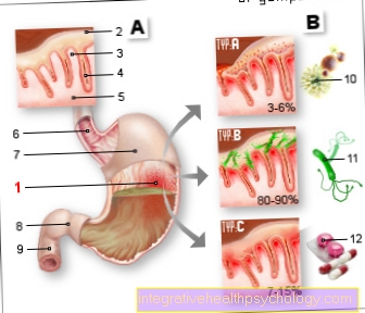
Illustration esophageal cancer

The tumor has already occluded a large part of the esophageal diameter.
This results in difficulty swallowing.
Sometimes food components can no longer pass through the constriction (stenosis).
Epidemiology
The incidence peak of esophageal tumors is between the ages of 50 and 60, with men 2-3 times more likely to be affected by this disease than women.
Overall, esophageal carcinoma is a comparatively rare cancer with an overall incidence of 10 cases per 100,000 population.
In Europe, esophageal carcinoma is represented among the total cancer deaths with 3.3% in men and 1.4% in women. However, women are more often affected by esophageal cancer that is higher up (close to the mouth), which in turn has an even worse prognosis than that stomach closer.
Frequency distribution within the esophagus:
- In the neck part (cervical) tumors located in the esophagus 5-10%
- Above the fork of the windpipe located tumors (suprabifurcal) 45-55%
- Tumors located below the fork of the windpipe (infrabifurcal)
- 40-50%
Anatomy of the esophagus

Illustration of the esophagus from the larynx to the diaphragm / stomach
- Cricoid cartilage
- Aortic narrowing (end of the abdominal artery)
- Zwerfellenge
- thyroid
- Carotid artery (carotid artery)
- Windpipe (trachea)
- right main brochius (bronchi)
- esophagus
- Diaphragm
Read more about anatomy under our topic: Anatomy of the esophagus

- esophagus
(Neck section) -
Esophagus, pars cervicalis - Nasal cavity - Cavitas nasi
- Oral cavity - Cavitas oris
- Windpipe (approx. 20 cm) - Trachea
- esophagus
(Chest section) -
Esophagus, pars thoracica - esophagus
(Abdominal section) -
Esophagus, pars abdominalis - Stomach entrance -
Cardia - Stomach body -
Corpus gastricum - Throat -
Pharynx - Thyroid -
Glandula thyroidea
You can find an overview of all Dr-Gumpert images at: medical illustrations
Forms and causes
Different forms of esophageal cancer and their causes:
Esophageal tumors generally occur primarily in the physiologically existing constrictions of the esophagus.
First of all, two main forms of this cancer will be distinguished:
In the upper part of the esophagus, the surface of the mucous membrane is mainly lined by squamous epithelium (covering tissue). The lower part consists mostly of glandular tissue.
Accordingly, depending on the origin of the cell type, squamous cell carcinomas mainly develop in the upper part of the esophagus and gland tumors (adenocarcinomas) in the lower part. More rarely, there is a special form of this cancer, which mainly grows lengthways along the wall. It destroys the autonomic nerves of the esophagus (plexus myentericus Auerbach), so that peristalsis is switched off and the esophagus is a rigid structure. This form is called hard (scirrhous) esophageal carcinoma.
- The main risk factors for squamous cell carcinoma (60%) are years of consumption of high-percentage alcohol (alcohol abuse). The cancer-causing (carcinogenic) effect of alcohol is increased by smoking cigarettes. These two pollutants are often mentioned in the same breath in relation to esophageal cancer, because in most cases they are consumed together.
For more information on this topic, we recommend our page on: Squamous Cell Carcinoma - How Dangerous Is It?
- In most cases, adenocarcinoma (40%) arises from a beret esophagus, a change in the mucous membrane that can develop after a long-standing reflux disease (chronic heartburn). It is therefore also called "beret carcinoma". It should be noted that by far not every beret esophagus inevitably develops into a tumor. In the last few decades there has been an increase in the secondary diseases caused by reflux disease and thus also in adenocarcinoma of the esophagus.
Other pollutants from food:
Carcinogenic substances are also found in food.There are chemical compounds made of nitrite (saltpeter, curing salt) and certain proteins (amines), so-called nitrosamines. Nitrosamines arise, among other things, when grilling, roasting and sometimes they arise in the stomach from foods that are particularly rich in nitrates such as Spinach or lettuce.
Aflatoxins are pollutants that are formed by certain molds in food. These can not only cause tumors in the esophagus but also in other organs, e.g. in the liver.
Similar effects are ascribed to milk mold, which preferentially affects dairy products.
Since moldy food is seldom consumed in Europe now, this cause of cancer is more widespread in the “third world countries”. In some regions of the world, betel nuts are chewed as a luxury food by large parts of the population. This pollutant can cause a wide variety of cancers, particularly in the mouth and esophagus.
After causticizing the esophagus with acids or alkalis, esophageal cancer can develop as a late consequence of the damage to the mucous membranes.
In the long run, hot drinks and spicy foods also cause similar irritation of the mucous membranes and can promote the development of a tumor. In some Asian countries, for example, a connection between the consumption of hot drinks and food and the occurrence of esophageal cancer has been established.
Vitamin deficiencies and poor hygiene are also discussed as the cause of the regional differences.
Diseases that can result in esophageal cancer:
Reflux disease (chronic heartburn) as the cause of tumor development has already been reported above.
A delayed passage of food is irritating to the mucous membrane. In diseases that delay the passage, the risk of developing an esophageal tumor increases. These include diseases such as achalasia and esophageal diverticula.
Achalasia is a widening of the esophagus in front of the stomach entrance. In the case of an esophageal diverticulum, there is a lateral bulging of the esophageal wall.
In particular, scars of the mucous membrane caused by caustic chemical burns can narrow (stenose) the esophagus, so that many years later a carcinoma can develop on the base of this scar.
After radiation exposure that affected the esophagus a long time ago, the risk of developing esophageal cancer increases due to the radiation damage.
The Plummer-Vinson syndrome describes a change in the mucous membrane (mucosal atrophy) in the area of the mouth, throat and esophagus.
The cause of this syndrome is a long-standing, chronic iron deficiency, which tends to occur in advanced age. The syndrome increases the risk of developing an esophageal tumor.
In some cases a family history of this cancer can be traced back. Genetic inheritance plays an important role here.
Symptoms
Symptoms in patients who have cancer of the esophagus, for example Difficulty swallowing, Pain when swallowing, hoarseness, frequent occurrence of to cough and Weight loss be.
Swallowing disorders are the most typical of the disease, but they usually only appear at an advanced stage.
Read more on the topic: Symptoms of esophageal cancer
Symptoms of early esophageal cancer
Esophageal cancer is a condition that occurs in most cases no complaints in the early stages caused. That makes it a very insidious disease.
Unfortunately, this is not infrequently the case with cancer in general. An early diagnosis is esophageal cancer almost always an incidental finding. Symptoms such as swallowing difficulties and hoarseness often only appear in the very advanced stages of the disease.
Signs of esophageal cancer
Cancers of the esophagus are among the diseases that often only cause symptoms at an advanced stage. This is particularly devastating with regard to the chances of recovery.
The leading symptom of esophageal cancer is one Swallowing disorder (Dysphagia). This can show up in different ways. For example, initially through a Feeling of pressure or Burning behind the breastbone when eating or feeling that the food is getting stuck.
In advanced stages, the uptake of liquid substances can become a problem due to the increasing narrowing of the esophagus by the tumor. Hoarseness is also a symptom that patients with esophageal cancer complain more often.
As with many other cancers, it also plays a role in cancer of the esophagus Weight loss as a symptom a not insignificant role.
A symptom complex that is described as "B symptoms" includes typical non-specific symptoms that often occur in the context of cancer: A unwanted weight loss of at least 10% of the original body weight within 6 months, otherwise unexplainable Fever over 38 ° C and profuse night sweats that makes it necessary to change clothes.
These B symptoms do not only occur in cancer, but also in infectious diseases such as tuberculosis. By no means all patients suffering from a malignant tumor disease show this complex of symptoms; it occurs relatively frequently in patients suffering from lymph gland cancer.
diagnosis
The most important examination for diagnosing esophageal cancer is the Reflection of the esophagus, Stomach and duodenum (Esophagogastroduodenoscopy).
Here, either after numbing the throat using a local anesthetic spray or after administering a sleep injection, a tube is pushed through the mouth and throat into the esophagus, stomach and duodenum. A camera is attached to the hose. With the help of this one can look at the organs.
If one area is noticeable, this one can small tissue sample (Biopsy). This is sent for a tissue examination. In this case, the piece of fabric is, for example, under the microscope considered, the pathologist can then make the diagnosis. The suspicion that a malignant disease is present can often be expressed during the reflection based on the external appearance of the conspicuous area, but a reliable diagnosis is always only possible under the microscope.
Especially in the area of the stomach and duodenum, a simple ulcer can look very similar to a tumor. For further diagnosis, the Endosonography, a mixture of mirroring and ultrasound, are used. With the help of this, for example, the depth distribution into the surrounding tissue can be assessed. This is often important in order to decide which therapy options are possible.
That is also important Search for tumor deposits. This is usually done using a Computed Tomography. Possible metastatic sites of esophageal carcinoma are primarily lymph nodes, lungs and liver.
therapy
Conservative therapy means non-invasive therapy, so there is no surgical intervention.
The most conservative therapeutic options for esophageal cancer include radiotherapy (Radiotherapy) and the chemotherapy or a combination of both.
Which Type of therapy is useddepends largely on the Tumor stage now and then how old the patient is and especially in which health condition he is located.
Sole application of radiation or chemotherapy without subsequent or previous surgery often only takes place in one palliative treatment instead of.
Palliative means that a cure is no longer possible complaints but as best as possible contained should be. Radiation and chemotherapy can be used to try to inhibit or slow down the growth of the tumor.
A newer method is the so-called photodynamic therapy. Here, the patient is administered a substance which accumulates relatively selectively in the tumor tissue. Subsequently, the tumor tissue with light of a certain wavelength irradiated. This leads to a so-called phototoxic reaction, some of the tumor cells are destroyed. This is used in the esophagus, for example, to somewhat reduce severe constrictions and thereby improve the passage of food again.
A Combination of radiation and chemotherapyHowever, so-called chemoradiotherapy is not only used in a palliative situation. In some cases it can be helpful to shrink the tumor using a combination of radiation and chemotherapy before surgery so that the surgery is more promising. One then speaks of neoadjuvant radiochemotherapy.
Another conservative therapy option is that Insertion of a metal tube (Stent) into the esophagus. This therapy also only serves that Relief from Symptoms and not healing. The stent can push the tumor mass to the edge a little and thus make swallowing a little easier again.
Surgical treatment of esophageal cancer
When a Operated esophageal cancer depends on the stage of the cancer, the age of the patient, and the overall condition of the patient.
Depending on which one height in the esophagus of the tumor sit, come various operations in question.
The esophagus runs through the chest and down into the upper abdomen. The tumor sits far below, so only has to Opened abdominal cavity become. However, it is not uncommon for a so-called 2-cavity procedure to be necessary, so the chest and abdominal cavity must be opened in order to be able to remove the tumor.
Is the tumor on Transition from the esophagus to the stomach, can a additional partial distance of Stomach be necessary. Partial or complete removal of the esophagus is usually necessary. In most cases a so-called Gastric elevation respectively. This means, as the name suggests, that the stomach is pulled up from the abdomen and made a kind of tube. It then serves as a Esophageal replacement. If the stomach is not an option as an esophageal replacement, the surgeon uses part of the large or small intestine, which he then installs between the stomach and the remains of the esophagus.
It is not uncommon a combination of radiation and chemotherapy before surgerywho have applied chemoradiotherapy. This allows a Shrinking the tumor which increases the chances of being able to completely remove the tumor with the operation.
For some years now, tumors have been diagnosed at a very early stage using a purely endoscopic procedure, i.e. within the framework of a Gastroscopy, remove. Here is the Tumor tissue with an electric snare “scraped off” the mucous membrane.
Risks The operation can include bleeding, infection with germs, allergic reaction to the anesthetic, injuries from the surgical instruments, injuries to neighboring organs and damage to nerves.
Complications
If the tumor is very advanced, its space-demanding (invasive) growth (Infiltration) in the Windpipe (trachea) grow into it. This can sometimes create an open connection between the two hollow organs, a so-called esophago-tracheal fistula. Through this fistula food components can get into the lungs and so repeatedly (recurrent) severe pneumonia cause. Especially under one radiotherapy the tumor can literally melt down and form fistulas.
In the case of esophageal ca, it can also be smaller chronic bleeding come, some of which lead to a relevant blood loss unnoticed, and such Anemia (Anemia) can cause. If the tumor is bleeding too much, you may vomit blood (Hematemesis) come.
metastasis
One can have two forms of Metastasis (tumor spread) describe:
- Lymphogenic metastasis:
The lymph vessels drain the Lymph fluid from all parts of our body and thus also from an esophageal tumor. Once this has connected to a lymphatic vessel through its growth, it happens that some tumor cells detach from the tumor cell cluster and are carried away with the lymph flow. Lymph nodes lie in the course of a lymph vessel. As a seat of the immune defense, they have the task of catching and fighting germs (bacteria). The tumor cells settle in the nearest lymph nodes and multiply again.
This creates a lymph node metastasis. This form of metastasis is the most common form in this type of cancer.
- Haematogenic metastasis:
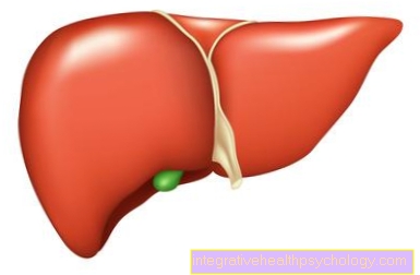
If the tumor becomes attached to a blood vessel as it grows, cells can tear themselves away, as in lymphogenic metastasis, and be dispersed throughout the body via the bloodstream. Most often, the tumor cells settle in the liver, lungs, brain and ribs and form what are known as distant metastases.
Life expectancy in esophageal cancer
The Life expectancy in patients with esophageal cancer is usually short. This is particularly because the cancer often occurs recognized late becomes.
Overall, the 5 year survival rate, i.e. the number of patients who are still alive 5 years after diagnosis, under 20%.
Will the Diagnosis at an early stage and if the tumor can be removed completely, the improvement will be forecast clear. Studies have found an average survival time of 9 months after diagnosis. However, this is an average value, so all stages, including the final stages, are included here. Patients with early-stage esophageal cancer often have a significantly longer life expectancy.