Thyroid scintigraphy
definition
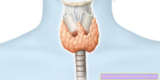
The thyroid scintigraphy is a radiological (more precisely: nuclear medicine) examination to diagnose the function of the organ. In contrast to ultrasound or cross-sectional imaging, it is not the structure that is shown, but the activity and thus the hormone production. To do this, a substance is added to the blood that accumulates in the thyroid gland and emits radioactive radiation. This can be measured using a special camera and converted into an image by a computer.
Indications
Thyroid scintigraphy is performed, for example, if lumps are found during the palpation examination or in the ultrasound image. In this way it can be examined whether these are hormone-producing or not. All knots from a size of 1cm should be clarified. If you have hyperthyroidism, one or more areas of increased activity may be the cause of the scintigraphy. A scintigraphy is also carried out, for example 6 months after radioiodine therapy (removal of diseased tissue by irradiation from the inside), to check whether the treatment was successful.
Scintigraphy for Hashimoto's thyroiditis
Thyroid scintigraphy is not common in the autoimmune disease Hashimoto. The determination of thyroid antibodies in the blood is particularly useful for diagnosis. In Hashimoto's disease, scintigraphy is most likely to show decreased activity of the entire thyroid gland.
preparation
A special preparation is usually not necessary for the thyroid scintigraphy. Anyone who takes medication that has an influence on the thyroid function should inform the examining doctor at the first examination, as this can influence the scintigraphy results. These include thyroid hormones (e.g. thyroxine), iodine tablets, amiodaraon (heart drug), or drugs that inhibit thyroid function (e.g. carbimazole). If necessary, these should also be discontinued a few days before the scintigraphy. In some cases the examination is carried out specifically under the influence of thyroid hormones, which are taken as tablets. This preparation usually takes place over a period of two to four weeks and the doctor will inform the patient accordingly in good time.
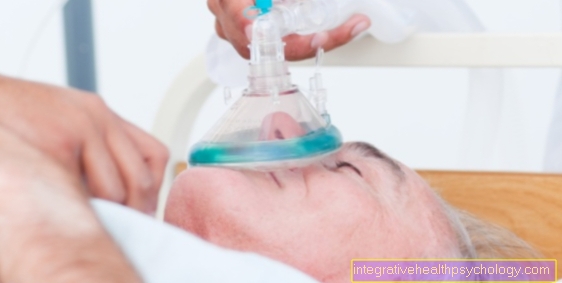
procedure
Thyroid scintigraphy can be performed on an outpatient basis in a radiological practice or in the thyroid outpatient department of a radiological clinic. It is not necessary to be admitted to the hospital for the examination.
First, the doctor injects a liquid that contains the radioactive substance into a vein, usually on the arm. Radioactive iodine or substances similar to iodine such as pertechnetate (radioactive element: technetium) are used, which are built into the thyroid just like iodine. You now have to wait about ten to twenty minutes. During this time, the radioactive particles are distributed with the blood in the body and thus also reach the thyroid. Almost exclusively there, some of them are recorded. Now the actual measurement is made by the so-called gamma camera, which you usually sit in front of. This registers the radioactive radiation (gamma radiation) that is now emanating from the thyroid gland. If a patient cannot sit down, scintigraphy is performed lying down. With the help of a computer calculation, an image corresponding to the distribution of the radiation is created. It also measures the amount of radiation that is administered and absorbed by the thyroid gland. This is the so-called "uptake".
The measurement itself takes about ten minutes and does not cause pain, nausea or other discomfort. The result is usually immediately available to the doctor and he can make initial statements. A report with all the information and the next steps will be sent to the patient and the family doctor soon. You can go home after the examination. However, contact with pregnant or breastfeeding women and children should be avoided for a few hours, or at least a distance should be kept as the body still emits some radiation. However, this breaks down continuously and is also excreted in the urine.
Evaluation / values
The evaluation of the scintigraphy of the thyroid is first carried out on the basis of the created image. All areas of the butterfly-shaped organ are shown in different colors. Blue tones stand for low tissue activity and red tones for high tissue activity. Areas with increased or decreased activity can therefore be determined with the optical assessment alone.
The second important aspect in the evaluation are the scintigraphy values, which are usually given as TcTU (technetium thyroidaler uptake = technetium uptake of the thyroid) in percent. This is the amount of radioactivity given by the syringe (in the form of technetium) that was ultimately absorbed by the thyroid gland. Usually the value is below 2%. It helps the nuclear medicine doctor assess a possible disease in conjunction with the other findings.
cancer
Thyroid scintigraphy cannot determine whether cancer is present. It can only give hints.
For example, if a thyroid nodule that is palpable or detected on ultrasound shows only weak activity on scintigraphy (cold nodule), it may be a cancerous tumor. In order to gain information, a so-called fine needle biopsy is usually recommended. Examination of the cells obtained may or may not corroborate the suspicion. The radiation exposure from scintigraphy of the thyroid itself is too low to pose a serious risk for the development of cancer.
Cold knot
A cold lump in the thyroid gland is when one area of the thyroid gland does not absorb any or at least significantly less radioactivity than the rest of the thyroid gland during the scintigraphy.
In the image (scintigram) created during the scintigraphy, this is usually shown as an area that contrasts with the rest of the thyroid in color. Accordingly, it is tissue that does not produce thyroid hormones. For example, it could be a harmless water-filled cyst. However, as thyroid cancer may be present in some cases, cold nodules should be examined by sampling to be safe.
A so-called fine needle biopsy is carried out for this purpose. Under local anesthesia and under visual control by an ultrasound head, the doctor takes a tissue sample from the knot with a long needle. If abnormal cells are found, surgical removal of the thyroid is usually recommended. The name of the cold nodule does not come from a temperature difference, but from its representation in the scintigraphy. Weak radioactivity is usually shown in blue.
Hot knot
If an area with stronger radioactivity stands out from the rest of the thyroid tissue in the scintigraphy, this is also known as a hot nodule. The higher the radioactive radiation, the redder the lump is shown. That justifies the naming and not an actual temperature difference.
There is also no connection to a possible inflammation. Hot knots represent areas of the thyroid gland with increased activity, that is, increased thyroid hormone production. These are so-called autonomous nodes or focal autonomies. These are areas that produce excessive hormones regardless of the body's control mechanisms. If these are particularly active, signs of hyperfunction such as tremors, palpitations, restlessness and many more can appear.
They can be cured by surgery or radiation therapy with radioactive iodine (radioiodine therapy). A malignant disease is rather unlikely with hot lumps unless there are also cold lumps. In the case of hot lumps, fine needle biopsy (sampling to examine for changed cells) is therefore usually not appropriate.
Risks
Thyroid scintigraphy is a very low-risk test. The radiation exposure is quite low.
Only pregnant women are at risk, as the child can develop deformities. Therefore, pregnancy speaks against scintigraphy. There is no danger for people with a so-called iodine allergy. It is an allergy that is directed not to iodine but to other components of contrast media containing iodine. However, these are not used in a scintigraphy.
Duration
The scintigraphy of the thyroid gland usually takes no longer than half an hour from the injection of the radioactive substance to the completion of the actual measurement. After completing the measurement, it must be ensured that radioactive radiation is still emitted for a few hours. Close contact with pregnant women, breastfeeding women and children should be avoided during this time. By the next day at the latest, the radioactivity has decayed and the substances excreted in the urine so that there is no longer any danger to fellow human beings.
Carbimazole
Carbimazole is a drug that inhibits the function of the thyroid gland and thus inhibits hormone production. It is used in the event of overfunction. Because of its influence on thyroid function, it also affects the results of a scintigraphy. Therefore it should be stopped a few days before the examination if possible. If the examination is nevertheless carried out under the influence of carbimazole, this must be taken into account in the evaluation.
Radiation exposure
Many people fear a thyroid scintigraphy because of the radioactive radiation used. The fear is largely unjustified, as one is only exposed to a very low level of radiation during this examination.
Our bodies experience low levels of radiation in everyday life anyway. In some situations it is higher, such as a long-haul flight. There are also regional differences. The additional radiation exposure of a scintigraphy of the thyroid is roughly equivalent to a natural radiation exposure of six months. If there is an indication for the examination, the advantages outweigh the low risks.
There is an exception for pregnant women, as the possible consequences of radiation exposure for the growing child would be particularly fatal. Thyroid scintigraphy should therefore not be performed during pregnancy. In children, the advantages and disadvantages must be weighed, but in principle a scintigraphy can be carried out with doses adapted to age and weight.


.jpg)