Lower jaw
introduction
The human jaw comprises two parts, the upper jaw and the lower jaw.
These two bony structures differ significantly in both size and shape. While the upper jaw (lat. Maxilla) is formed by a pair of bones and is firmly connected to the skull bone, the lower jaw (lat. Mandible) consists of a very large, compact portion of bone and comes into free contact with the skull via the two temporomandibular joints.
For this reason, the lower jaw forms the movable part of the jaw that is essential for the chewing process. A general distinction is made between two large parts of the skull, the Facial- and the Brain skull. Those parts of the bone that wrap around the brain like a shell and thus fulfill a certain protective function are counted as part of the so-called brain skull.
The bones of the facial skull, in turn, define the basic features of the human face. The facial skull includes:
- Parts of the Frontal bone
- of Temporal bone
- the Nasal bone
- the Zygomatic bone
- the Tear bone
- the paired Turbinate
- the Ethmoid bone and
- the Ploughshare
The upper and lower jaw are also anatomically counted as part of the facial skull. In contrast to the upper jaw, the lower jaw hardly fulfills a protective function. It is flexibly connected to the rest of the skull via the temporomandibular joints and is mainly responsible for chewing and interacting with the tongue important for language education.
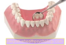
Figure lower jaw
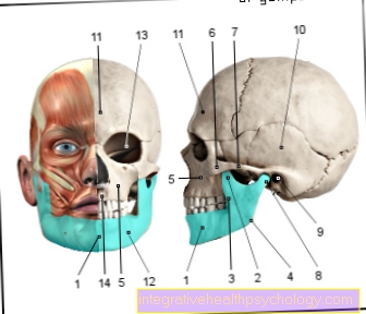
- Lower jaw - Mandible
- Crown process -
Coronoid process - Lower jaw rest -
Ramus mandibulae - Mandible angle -
Angulus mandibulae - Upper jaw - Maxilla
- Zygomatic bone - Os zygomaticum
- Zygomatic arch -
Arcus zygomaticus - Temporomandibular joint -
Articulatio temporomandibularis - External ear canal -
Meatus acousticus externus - Temporal bone - Temporal bone
- Frontal bone - Frontal bone
- Chin hole - Mental foramen
- Eye socket - Orbit
- Upper jaw, alveolar process -
Alveolar process
You can find an overview of all Dr-Gumpert images at: medical illustrations
Structure of the lower jaw
Of the Lower jaw (lat. Mandible) consists of a horseshoe-shaped bone structurewhose body (lat. Corpus mandibulae) forms. The front edge of the lower jaw forms the human chin. The large body of the lower jaw is raised on both sides by an ascending branch, the Lower jaw branch (lat. Ramus mandibulae) continued.
The body of the lower jaw and the ascending branches together form an angular structure, the Mandibular angle (lat. Angulus mandibulae), which as the approach and origin of various am Chewing process involved muscles. Basically, one differentiates between three processes on this bone of the facial skull.
On the upper side of the body of the maxilla is the Alveolar process (lat. Alveolar process), embedded in it are the alveoli, small indentations that accommodate the Tooth roots serve.
In the area of the ascending branch, another process, the so-called extension, goes off the bone Articular process (lat. Condylar process or Articular process). This in turn has a roller-like Swivel head on, which is the moving part of the Temporomandibular joint forms. The so-called Muscular process (lat. Muscular process) forms the point of attachment of various muscles.
A small protrusion can be seen on the inside of the lower ramus. This structure is used in the anatomy as Bone tongue (lat. Lingula mandibulae) designated. It covers a small hole that runs across the bone of the lower jaw (lat. Mandible foramen) and as the passage point for the Mandibular nerve (Inferior alveolar nerve) acts.
Supply lower jaw
The lower jaw is sensibly supplied by the large lower jaw nerve Inferior alveolar nerve. This nerve represents a splitting off of the Mandibular nerve which in turn emerges from the fifth cranial nerve, the trigeminal nerve. Both the supplying nerve, Inferior alveolar nerve, as well as the responsible vessels (Artery and Inferior alveolar vein) run through a canal located inside the maxillary bone. This channel (Mandibular Canal), runs like a tunnel under the teeth of the lower jaw, from there nerve fibers and splits of the vessels reach the individual teeth.
Read more about anesthesia in the lower jaw at: Conduction anesthesia at the dentist
Teeth-supporting apparatus
With the help of the so-called Teeth supporting apparatus is every single one tooth relatively firmly anchored in the lower jaw. In order to meet the demands of the purchase process and the various protective functions, the tooth support apparatus exists in both upper jaw as well as in Lower jaw from different proportions.
Deep indentations within the jawbone (lat. Alveoli) accommodate the Root part of every tooth. The tooth support system also includes:
- the superficially localized Gums (lat. Gingiva propria),
- the Dental cement (Cementum) and
- the Root skin (Periodontal disease or Periodontium).
However, if you take a closer look at the tooth support system, you can quickly see that the individual Teeth not absolutely tight and are rigidly fixed in the jawbone. Such anchoring would also be absolutely counterproductive in view of the forces that act on the teeth during the chewing process. In fact, every single tooth is through Collagen fiber bundles, the so-called Sharpey fibers suspended resiliently in the alveolus. As a result, the tooth remains relatively mobile and the forces and during the Chewing process resulting pressures can be effectively distributed over a larger area. The stress that acts on each individual tooth is reduced enormously in this way. Furthermore, the tension of these collagen fiber bundles during the chewing process prevents the Tooth roots Press too deep into the jawbone under the influence of pressure.
Diseases lower jaw
To the typical diseasesn that can occur in the area of the lower jaw belong Inflammation in the area of the teeth and bones. Furthermore are Broken bones the lower jaw is not uncommon, but can be treated relatively well. Other frequently occurring diseases of the lower jaw manifest themselves less in the area of the actual bone than in the area Temporomandibular joint.
Excessive and / or incorrect stresses on the joint can lead to a so-called Lock jaw or to Jaw clamp come.
Under the term Jaw clamp (Trism) one understands a restricted respectively incomplete mouth opening. In most cases there is one Spasm of the masticatory muscles cause of the occurrence of a clamp. Also can local inflammatory processes lead to the development of a jaw clamp in the area of the masticatory muscles. Affected patients report problems opening their mouths, the lower jaw is only minimally lowerable in the temporomandibular joint and can be lowered with severe pain. The jaw clamp as a disease of the lower jaw is caused by passive stretching exercises treated. This therapy method lasts several weeks in many cases and is very uncomfortable for the patient concerned.
As Lock jaw known disease of the lower jaw shows up in contrast as mfishing final ability of the jaw. The rows of teeth can therefore no longer be perfectly placed on top of one another. Possible causes for the occurrence of a locked jaw are Dislocations of the temporomandibular joint, Fractions in the area of the joint head and abnormal changes the bone structure. By far most common reason for the development of this clinical picture is one excessive mouth opening while yawning or biting an apple.