Causes of hip osteoarthritis
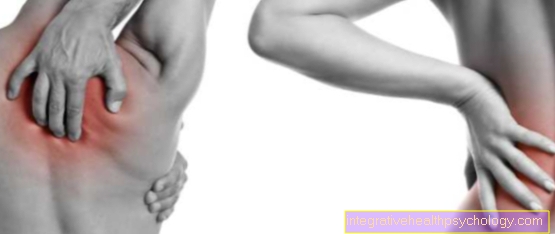
Hip pain
Are you looking for the cause of your hip pain or you don't know exactly what is causing your hip pain?
Then let yourself be guided by our diagnostic Hip pain guide and come to the most likely diagnosis.
Causes of the development of hip arthrosis
There are different causes for a Hip osteoarthritis.
In addition, the causes of hip osteoarthritis are in most cases not known. In order to better differentiate osteoarthritis with regard to its cause, a distinction is made between primary and the secondary osteoarthritis. If there is a arthrosis Developed without an apparent trigger - and this is the case in most cases - is called one primary osteoarthritis.
Secondary hip osteoarthritis, on the other hand, is arthrosis that is caused by previous damage, incorrect loading, individual inflammatory processes or incorrectly placed hip socket (Hip dysplasia) or Femoral neck (Impingement) arises.
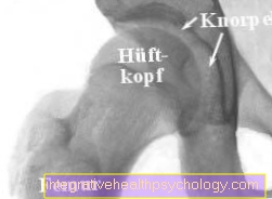
The x-ray in the top right shows you a healthy hip. You can see the distance between the femoral head and the acetabulum very well. This distance implies that both the socket and the femoral head are covered with a good layer of cartilage. This is no longer the case with osteoarthritis. In such a case, the cartilage layer shows considerable damage.
Listed below you will find the most common causes that can be responsible for the development of hip arthrosis. In most cases there is more information available to you that can be accessed via a link.
- Congenital partial or complete dislocation of the hip (hip dislocation) in congenital hip dysplasia:
In around 10% of all newborns, the femoral head is not positioned correctly in the socket. The diagnosis is usually made through an ultrasound scan. Depending on the individual extent of the hip dysplasia, treatment with a spreading bandage or an operation is necessary to prevent long-term effects. It is of particular importance here that this clinical picture is recognized as early as possible, as subsequent maturation of the acetabular roof (reduction or elimination of hip dysplasia) is only possible within the first two years of life. If there is no diagnosis or no treatment, permanent hip dysplasia develops with its long-term consequences of hip arthrosis. - Congenital form disorder (hip dysplasia):
One speaks of hip dysplasia in patients whose hip socket is too flat or whose femoral neck angle is too steep (see also the anatomy of the hip joint). As a result, the socket roof does not completely cover the femoral head, which means that the load is only borne by too small a part of the joint.
This leads to premature wear of the hip joint. It therefore seems plausible that such so-called pre-arthrotic changes (= malpositions causing osteoarthritis) should be surgically corrected at an early stage if the findings are severe. This can be done, for example, by so-called adjustment osteotomies.
When comparing the x-ray image of hip dysplasia with the x-ray image of a healthy hip (see above), serious differences can be seen. It seems logical that this cannot be without consequences. In a gender-specific comparison, it is noticeable that women suffer relatively more often from hip dysplasia. The ratio between women / men is around 9: 1. - Metabolic disorders:
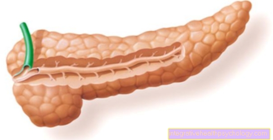
Diabetes mellitus
Diabetes mellitus causes changes in the blood vessels, which in turn lead to circulatory disorders in the area of the femoral head. The consequence of this insufficient supply is, for example, a deformity of the femoral head or, in the worst case, the death of the femoral head (femoral head necrosis, see below).
You can find more information under our topic: Diabetes mellitus
Appointment with a hip expert?

I would be happy to advise you!
Who am I?
My name is dr. Nicolas Gumpert. I am a specialist in orthopedics and the founder of .
Various television programs and print media report regularly about my work. On HR television you can see me every 6 weeks live on "Hallo Hessen".
But now enough is indicated ;-)
The hip joint is one of the joints that are exposed to the greatest stress.
The treatment of the hip (e.g. hip arthrosis, hip impingement, etc.) therefore requires a lot of experience.
I treat all hip diseases with a focus on conservative methods.
The aim of any treatment is treatment without surgery.
Which therapy achieves the best results in the long term can only be determined after looking at all of the information (Examination, X-ray, ultrasound, MRI, etc.) be assessed.
You can find me in:
- Lumedis - your orthopedic surgeon
Kaiserstrasse 14
60311 Frankfurt am Main
Directly to the online appointment arrangement
Unfortunately, it is currently only possible to make an appointment with private health insurers. I hope for your understanding!
Further information about myself can be found at Dr. Nicolas Gumpert
- Gout:
Patients with gout have high levels of uric acid in their blood. If the uric acid content is around 8 mg / dl or higher, there is a likelihood of so-called uric acid crystals (= Urate crystals) in the joint most likely. These crystals destroy the actually smooth surface of the joint. Crystals are deposited when the level of uric acid in the blood is too high, which can lead to a gout attack.
For more information, see our topic: Gout
- bacterial coxitis:
This includes infections of the hip joint caused by bacteria, which can lead to hip osteoarthritis. The appearance of a bacterial Coxitis is theoretically possible for all people, but the likelihood of developing bacterial coxitis is significantly increased, especially in children and patients with an artificial hip joint.
In children, for example, it can arise when a focus of infection is spread via the bloodstream. For example, a Tonsillitis bacterial koxitits or chronic bone suppuration by spreading via the bloodstream (Osteomyelitis) cause.
Please also read the topic osteomyelitis.
Epiphyseal capitis femoris
(in children and adolescents, in boys between around 12 and 16 years of age, in girls usually between 10 and 14 years of age):
In the child, the femoral head is formed by the so-called growth plate (= Cartilage joint) Cut. This means that the femoral head and the femoral neck are not yet continuously bony connected. If loosening occurs in the growth plate between the femoral neck and head, this can result in separation and displacement. This means that when the load is placed on the femoral neck upwards (= ventrally cranial) migrates, but the femoral head is held in the acetabulum. An acute epiphyseal solution is always one of the few emergencies in orthopedics.That means: The epiphysis must be reduced as soon as possible. This means that the hip head that has slipped off must be quickly returned to its original position.A surgical procedure with repositioning and fixation of the femoral head may be necessary. With early diagnosis and surgical treatment with appropriate correction, the diagnosis can be classified as good. Permanent damage cannot always be ruled out, especially in the case of a late diagnosis. This means that if there is also femoral head necrosis, there is a risk of early secondary coxarthrosis. More information on this topic can be found at Epiphyseolysis capitis femoris.
- Joint chondromatosis
This is a conversion of mucous membrane tissue into cartilage cells that form cartilage balls, which as free joint bodies disrupt the mechanics of the joint and can lead to hip arthrosis.
- Femoral head necrosis (HCN):
Femoral head necrosis is a local circulatory disorder in the femoral head. The circulatory disorder leads to a deformation of the femoral head. The subsequent incongruence of the femoral head and acetabulum leads to the development of hip arthrosis for a short time.
- Perthes disease:
Circulatory disorders of the child's femoral head with increasing deformation of the femoral head. Perthes disease is comparable to femoral head necrosis. But since it occurs during the growth phase, incongruities can be compensated for by the still existing growth.
- Osteoradionecrosis:
This means the death of the femoral head due to circulatory disorders (femoral head necrosis) as a result of irradiation of an area near the hip joint in tumor therapy.
- Protrusio acetabuli:
This refers to the bulging of the hip socket into the pelvis. This disease is found more often in diseases of the rheumatic type (rheumatoid arthritis / rheumatism).
- Rheumatoid arthritis (rheumatism, chronic polyarthritis):
Rheumatoid arthritis starts on the inside of the skin Joints (= Synovia). The chronic inflammatory process releases substances that attack and ultimately destroy your own joint. You can find more detailed information under the heading Rheumatism / Rheumatoid Arthritis.
Injuries to the hip joint or hip dislocation:
Bone fractures in the area of the acetabular socket (acetabular fracture) or the femoral neck (femoral neck fracture) and dislocations in the hip area after accidents.Read more on the subject Femoral neck fracture and Femoral neck fracture late effects