Forebrain
synonym
Prosencephalon
Definition forebrain
The forebrain is part of the brain and is therefore part of the central nervous system.
It includes the diencephalon (Diencephalon) and the cerebrum (Telencephalon).
These emerge from the forebrain vesicle in the embryonic development phase of the brain.
The forebrain has a variety of functions, the cerebrum is essential for numerous processes such as motor skills, vision, hearing and many others, the diencephalon, which includes the hypothalamus and the pituitary gland, plays a central role in the hormonal control loop.
Diencephalon
Synonym: Diencephalon
The Diencephalon borders down (caudal) to the midbrain (Mesencephalon) to the Brain stem is counted.
At the top it borders on that Cerebrum, although it is difficult to make a precise definition here.
The diencephalon consists of Thalamus, Epithalamus, Subthalamus and Hypothalamus.
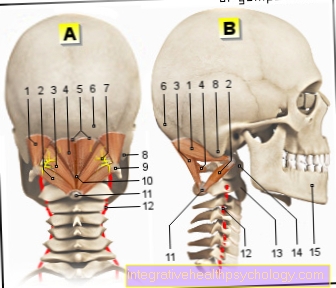
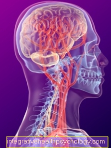
Illustration brain
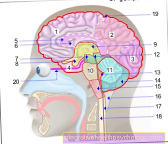
Cerebrum (1st - 6th) = endbrain -
Telencephalon (Cerembrum)
- Frontal lobe - Frontal lobe
- Parietal lobe - Parietal lobe
- Occipital lobe -
Occipital lobe - Temporal lobe -
Temporal lobe - Bar - Corpus callosum
- Lateral ventricle -
Lateral ventricle - Midbrain - Mesencephalon
Diencephalon (8th and 9th) -
Diencephalon - Pituitary gland - Hypophysis
- Third ventricle -
Ventriculus tertius - Bridge - Pons
- Cerebellum - Cerebellum
- Midbrain aquifer -
Aqueductus mesencephali - Fourth ventricle - Ventriculus quartus
- Cerebellar hemisphere - Hemispherium cerebelli
- Elongated Mark -
Myelencephalon (Medulla oblongata) - Big cistern -
Cisterna cerebellomedullaris posterior - Central canal (of the spinal cord) -
Central canal - Spinal cord - Medulla spinalis
- External cerebral water space -
Subarachnoid space
(leptomeningeum) - Optic nerve - Optic nerve
Forebrain (Prosencephalon)
= Cerebrum + diencephalon
(1.-6. + 8.-9.)
Hindbrain (Metencephalon)
= Bridge + cerebellum (10th + 11th)
Hindbrain (Rhombencephalon)
= Bridge + cerebellum + elongated medulla
(10. + 11. + 15)
Brain stem (Truncus encephali)
= Midbrain + bridge + elongated medulla
(7. + 10. + 15.)
You can find an overview of all Dr-Gumpert images at: medical illustrations
Thalamus
Anatomy & function:
Of the Thalamus is in pairs, i.e. on both sides, present and limited by its inner wall (medial) the third ventricle, one of several with brain water (Liquor) filled cavities of the brain.
The outer wall (lateral) of the thalamus is adjacent to the Internal capsule (inner capsule), through which numerous nerve fiber bundles en route to the Cerebrum or run from the cerebrum to the periphery.
The thalamus is made up of numerous nerve cell nuclei that are connected to one another by nerve fibers.
There are also numerous nerve fiber connections between the Thalamic nuclei and the Cerebrum.
Almost all sensory or sensitive Paths that run from the periphery into the cerebrum project first into the thalamus and from there into the cerebrum.
Therefore, the thalamus is sometimes also called "Gateway to the cerebral cortex" designated.
Many of the human sensations, such as sight (via the corpus geniculatum laterale) and listen (via the corpus geniculatum mediale), are integrated and presorted in the thalamus in order to avoid overstimulation of the cerebrum.
When they have been processed in the thalamus, they reach the cerebrum and are only then consciously perceived.
The thalamus also plays its part in processing motor impulses. So he gets information from the Cerebellum and the Basal gangliawhich play a crucial role in coordinating movements.
Furthermore, the thalamus plays an important role in the state of activity, i.e. tiredness or sleep as well as wakefulness and excitement and directed attention.
clinical evidence:
Damage to the thalamus can result in a variety of symptoms.
Usually a lesion of the thalamus is on one side mutual half of the body affected. This is due to the fact that almost all fibers that run from the periphery to the center or, vice versa, cross over to the opposite side.
Touching the right half of the body projects into the left half of the brain. A movement that is planned in the left cortex takes place in the end on the right half of the body.
Depending on the extent and localization of the damage to the thalamus, hemiplegia, impaired sensation, loss of vision on one side, restlessness of movement, pain without a recognizable pain stimulus and impaired consciousness can occur.
Epithamalus
The epithalamus sits on top of the thalamus from behind.
Two important structures of the epithalamus are the Epiphysis (pineal gland) and the Area pretectalis.
The epiphysis produces Melatonin. This is an important hormone in mediating the circadian rhythm and thus the sleep-wake rhythm.
The area pretectalis plays a role in the Interconnection of the pupillary reflex, i.e. the narrowing of the pupil when exposed to light.
She receives information from the Retina (retina) on the Optic nerve (Optic nerve) and sends nerve fibers to a nerve cell nucleus (Nucleus Edinger-Westphal) whose neurons are then used to activate the muscle that leads to the constriction of the pupil (Sphincter pupillae muscle), to lead.
It is important that the nerve fibers that are supposed to "report" the incidence of light not only to the Edinger-Westphal nucleus (Accessor nucleus of the oculomotor nerve) the side on which the light actually entered the eye, but also to the core of the opposite side.
The incidence of light that is registered in one eye therefore ultimately leads to Narrowing of the pupils in both eyes (consensual light reaction).
Subthalamus
Below the thalamus is the subthalamus.
Functionally it belongs to the Basal gangliathat is part of the Cerebrum are.
He thus plays a role in coordination and Fine-tune movements. The basal ganglia will be discussed in more detail below.
Hypothalamus
Below the subthalamus is the hypothalamus.
It forms the bottom of the 3. Ventricle (the thalamus forms its lateral limit).
It contains the Pituitary gland and leaves the Corpora mamillaria recognize those at the border from the hypothalamus to the Midbrain lie.
Also the Optic nerve, the optic nerve and second cranial nerve, as well as the optic nerve junction, that Optic chiasm, become evolutionary to Diencephalon counted.
The hypothalamus is, so to speak, the center or the highest station for the integration, processing and coordination of vegetative functions including the control of the endocrine organs, i.e. those organs that Hormones secrete.
The hypothalamus is jointly responsible for processes such as breathing, circulation, body temperature, fluid and food intake behavior, reproductive behavior, sleeping and waking (circadian rhythm) and many others. Some will be discussed in more detail here.
The hypothalamus contains different core groups, each of which has its own function.
An important part of the hypothalamus is that Pituitary gland. It lies - limited to the bones - in the Sella turcica (Turkish saddle), which borders the sphenoid sinus. This is the reason why surgical interventions on the pituitary gland usually over the nose be performed.
The pituitary gland is divided into two parts.
Of the Posterior pituitary lobe (Neurohypophysis) and the Anterior pituitary gland (Adenohypophysis), which is not part of the central nervous system is. It does not consist of nerve tissue but of glandular tissue and, in the strict sense, does not belong to the hypothalamus.
The neurohypophysis produces the hormones Vasopressin (also known as antidiuretic hormone = ADH) and Oxytocin.
Vasopressin plays a crucial role in the reabsorption of water in the kidney, moreover, it leads to a Vasoconstriction (Vasoconstriction).
This hormone is released when the hypothalamus registers that the body has too little water. In addition, a feeling of thirst is triggered, so that more water is supplied by drinking.
Oxytocin is an important hormone in pregnant, giving birth and breastfeeding women. So it takes care of one Contraction of the uterus, a labor induction during childbirth, and is used for breastfeeding Milk penetration responsible.
The anterior lobe of the pituitary gland lies just below the optic nerve junction (Optic chiasm), so that Tumors of the pituitary gland can lead to visual field defects.
The adenohypophysis produces hormones that act on thyroid, Adrenal glands, Mammary gland, Testicles or Ovaries and growth have an immense impact.
Here they are controlled by the hypothalamus as the higher-level center.
So the hypothalamus secretes hormones, which in turn ensure that the pituitary gland produces and releases hormones or not.
The hormones of the thalamus, which have a stimulating or inhibiting effect on the hormone production of the adenohypophysis, are in the area of the Tuber cinereum, another part of the hypothalamus. they call themselves Releasing hormones and through their effect on the pituitary gland, among other things, they affect the thyroid and cortisol metabolism.
Also belonging to the hypothalamus Corpora mamillaria have numerous connections with the Hippocampus, they play such a role in behavior and memory retention.
clinical evidence:
Numerous pathologies can occur due to disorders in the area of the hypothalamus.
Two diseases are mentioned here as examples. The central one Diabetes insipidus occurs when the anterior pituitary gland is damaged. Then the hormone is missing ADH, which is normally used for reabsorption of water in the kidney cares.
As a result, those affected up to 20 liters of urine per day to leave (Polyuria) and suffer from constant strong thirst and drink large amounts (Polydypsia).
Another form of the Diabetes insipidus is the renal (so caused by the kidney) Shape. The anterior pituitary lobe is sufficient here ADH who produces kidney but there is a lack of receptors that recognize and bind the hormone. Thus, ADH cannot develop its effect.
A destruction of the Corpora mamillariahow they especially by chronic alcohol abuse occurs, leads to clear behavioral problems and pronounced memory impairment.
Cerebrum
Synonym: Telencephalon
Definition:
The Cerebrum is also known as the endbrain and is part of the central nervous system.
It consists of two halves of the brain (Hemispheres), which are separated from each other by the fissura longitudinalis cerebri.
The two hemispheres can still be in four lobes subdivide.
Countless integration processes take place here, including the following:
- Motor skills
- See
- Listen
- Feel
- behavior
- memory
Anatomy:
A cerebral hemisphere is made up of four lobes:
- Frontal lobes
- Temporal lobe
- Parietal lobes
- Occipital lobe
None of these four areas could be assigned to the Cingulate gyrus which runs above the cerebral bar and the insula or island cortex.
The surface of the brain is heavily folded and thus of turns (Gyri) and furrows (Sulci) crossed. This results in an extensive increase in the surface area.
According to histology, the cerebrum can be seen in 52 different bark fields classify, these are named after their first description Brodman areas designated.
They are also part of the cerebrum Basal ganglia. They lie in the medullary bed, i.e. below or further inside than the cerebral cortex (subcortical). They play a central role in coordinating and fine-tuning movements.
Basal ganglia
Anatomy & function:
To the Basal ganglia counting Striatum - consisting of Caudate nucleus and Putamen – Pallidum, Subthalamic nucleus and Substantia nigra.
The subthalamic nucleus is actually located in the subthalamus, part of the Diencephalon. Functionally, however, it belongs to the basal ganglia.
Adjacent to the area of the basal ganglia is the Internal capsulethrough which numerous nerve fibers run in a central or peripheral direction. It borders the thalamus.
The basal ganglia are with each other and with the cerebral cortex (Cortex) closely networked via numerous nerve fibers.
They work as a complex network. They inhibit or activate each other in complex control loops and thus ensure a Fine tuning of motor skillswhich is first roughly planned by the cortex.
clinical evidence:
Lesions in the area of the basal ganglia can lead to diseases that result in motor disorders.
For example that Parkinson's disease. This is characterized by a lack of mobility (Akinesia), a rigor (increased muscle tone with stiffness of the muscles) and a resting tremor.
The cause is a lack of the messenger substance Dopamine in the field of Substantia nigra accepted.
A nearly opposite clinical picture is that Chorea huntington. It impresses - among other symptoms - through excessive movements of the extremities and the facial muscles.
There is a degeneration of nerve cells in her Striatum underlying.
Olfactory brain
Synonym: olfactory cortex
Anatomy & function:
The olfactory brain is located in the area of the paleocortex, the historically oldest part of the cerebral cortex.
It is found in the lower area of the frontal lobe (so frontobasal).
The first stage in the development of odor perception is Sensory cells of the olfactory mucosa. Your nerve cell extensions form the Olfactory nerve, the first of the twelve cranial nerves.
This runs to the one located in the frontal lobe Olfactory bulb. From there the nerve fibers pull over the Olfactory tract up to the olfactory cortex.
From here, the information reaches numerous other places, including via the thalamus into the neocortex, where olfactory perception is analyzed, interpreted and finally recognized and into the Amygdala (Almond kernel).
Limbic system
Anatomy & function:
The centers belonging to the limbic system are in some cases not very clearly defined. They are all close to the Cerebral bar (Corpus callosum).
The following structures are usually included in the limbic system:
- the hippocampus
- the amygdala
- the cingulate gyrus
- the parahippocampal gyrus
- the corpora mamillaria
The amygdala is located in the temporal lobe. It plays a decisive role in the emotionally conditioned regulation of vegetative parameters. For example, it helps our heart beat faster when we are frightened.
This is possible through numerous fiber connections in the amygdala to centers of vegetative regulation in the Brain stem and Hypothalamus.
It is also decisively involved in the control of fear and anger behavior, the emotional evaluation and recognition of situations and the connection, for example, of a smell or something heard with a certain emotion.
Of the Hippocampus Like the amygdala, it lies in the temporal lobe. He too has a share in vegetative and emotional processes.
However, it is more popular for its memory function. The so-called Papez neuron circle a crucial role.
From the hippocamous fibers pull in the Fornix up to the Corpora mamillaria of the hypothalamus. From there, the fibers continue to run over the Thalamus in the Cingulate gyrus, further into the Parahyippocampal gyrus and back to the hippocampus so that the neuron circle closes.
This complex network of nerve fibers is essential for the adequate functioning of the Short term memory indispensable.
clinical evidence:
Even the destruction of just one of the members of the Papez neuron circuit leads to massive memory impairment.
This affects newly learned content that cannot be kept for longer than one or two minutes.
Old memory contents, on the other hand, remain untouched because they have already been transferred from short to long-term memory.
Neocortex
Synonym: Isocortex
The neocortex is evolutionary youngest part of the brain.
It consists of four lobes:
- Frontal lobes
- Parietal lobes
- Occipital lobe
- Temporal lobe
Histologically, it consists of 6 cell layers.
A detailed description of the brain lobes can be found here: Neocortex