White matter spinal cord
Synonyms
Medical: Substantia alba spinalis
CNS, spinal cord, brain, nerve cell, gray matter spinal cord
English: spinal cord
Spinal cord in general
The Spinal cord Like the brain, it belongs to the central nervous system (CNS) and runs in the spine, more precisely in the spinal canal.
The spinal cord joins part of the brain at the top, the medulla oblongata (elongated marrow), which leaves the skull through a bony hole, the so-called foramen magnum.
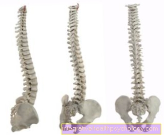
The spinal cord runs protected in the Spinal canalformed by the vertebral bodies stacked on top of each other. The spinal cord runs up to about the level of the first or second lumbar vertebrae. Down runs the spinal cord in the so-called conus medullaris (medullary cone) pointed to. This ends in the filum terminale, many thin connective tissue fibers. Nerve fibers of the lower spinal nerves, which are called cauda equina (horse's tail) due to their morphology, are found below the second lumbar vertebra.
White matter of the spinal cord
The white matter of the spinal cord consists of the predominantly myelinated nerve fibers that ascend and descend (the nerve fibers that are dosed and isolated by a fat sheath).
These are bundled as different strands (funiculi) in which they are each subdivided into tracts or fasciculi (= "small bundles") with different functions.
The cell bodies (perikaryen) of the Nerve cell are either in the brain or in the Spinal cord:
If they are in the brain, they are called the train descending (efferent) because information flows from the brain to the spinal cord. If they are in the spinal cord, they are called the web ascending (afferent) because the information flows from the spinal cord to the brain.
Some of the ascending and descending fibers, however, form fibers that lead to the Own device of the spinal cord; they are called basic bundles (Fasciculi proprii = "own bundles").
They lie directly against the gray matter and convey information within the spinal cord.
You can roughly distinguish one on each side
- Fore strand (Anterior / ventral funiculus)
- Side strand (Funiculus dorsalis / lateralis)
- Back strand (Funiculus posterior or "mediales lemniscus system")
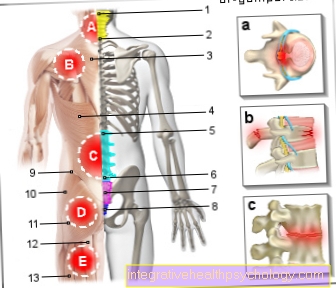
Figure spinal cord
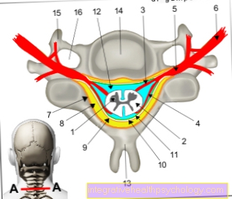
1st + 2nd spinal cord -
Medulla spinalis
- Gray matter of the spinal cord -
Substantia grisea - White spinal cord substance -
Substantia alba - Anterior root - Radix anterior
- Back root - Radix posterior
- Spinal ganglion -
Ganglion sensorium - Spinal nerve - N. spinalis
- Periosteum - Periosteum
- Epidural space -
Epidural space - Hard spinal cord skin -
Dura mater spinalis - Subdural gap -
Subdural space - Cobweb skin -
Arachnoid mater spinalis - Cerebral water space -
Subarachnoid space - Spinous process -
Spinous process - Vertebral bodies -
Vertebral foramen - Transverse process -
Costiform process - Transverse process hole -
Foramen transversarium
You can find an overview of all Dr-Gumpert images at: medical illustrations
Spinal tracts
Sensitive (= ascending, afferent) pathways:
Sensitive paths are used to process pulses / information from e.g. responsible for the skin and forwards this information to the appropriate centers in the brain.
- Spinobulbaris tract.
It leads fibers of tactile and depth sensitivity and consists of the- Fasciculus gracilis (GOLL) for the lower half of the body (lies inside) and the
- Fasciculus cuneatus (BURDACH) for the upper half of the body (lies on the outside)
For more information on the spinobulbaris tract, see our topic: Spinobulbaris tract
- Anterior and lateral spinothalamic tracts.
It directs pain, temperature, and gross pressure, including those in summary protopathic sensitivity, and is in the so-called Front strand, the anterolateral funiculus.
For more information on the spinobulbaris tract, see our topic: Tract spinothalamicus
The two lanes 1 and 2 each consist of 4 Neurons and pull both over the Thalamus, our "gateway to consciousness".
- Anterior spinocerebellar tract (GOWERS) and posterior (FLECHSIG) (= anterior and posterior cerebellar lateral cord tract)
It directs information from the muscle, joint and tendon spindles (Proprioception) to the Cerebellum (Cerebellum) and also lies in the anterior lateral cord (funiculus anterolateralis). This pathway consists of 3 nerve cells and does not run over the thalamus.
- Spinoolivaris tract (so-called triangular tract)
- Spinotectal tract
Motor spinal tracts
Motor pathways
Motorized paths are used for processing impulses / information from e.g. responsible for the skin and forwards this information to the appropriate centers in the brain.
- Corticospinal tract (pyramidal tract)
It conducts voluntary motor impulses from the cerebral cortex (= cortical) to the anterior horn cells in the spinal cord (spinalis). It is divided into two parts and lies in the anterior strand of the white spinal cord substance at the very side (corticospinal tract) and at the very front (corticospinal tract anterior).
- Extrapyramidal lanes
These are all tracks that do not belong to the “pyramid track”. They all have an origin below the cerebral cortex (subcortical) in different core areas of the brain and pull to the - or - motor neurons in the anterior horn of the spinal cord. They include the
- Reticulospinal tract:
These fiber bundles arise from the reticular format Brain stem and terminate at intermediate neurons in the gray matter of the spinal cord, which modulate the activity of the motor anterior horn cells. He provides i.a. the pathway between the respiratory center and motor neurons for the Respiratory muscles It runs in the lateral cord of the spinal cord, but is also scattered in the anterior cord.
- Vestibulospinal tract:
These fibers arise from the vestibular core group (Ncl. Vestibularis lateralis) in the hindbrain, which are more or less for our balance is responsible, and also influence the motor anterior horn cells via inter-neurons.
This happens because the intermediate neurons tell the motor anterior horn cells which muscles need to be tensed, which are needed to maintain our balance or a certain basic tension in the muscles (= tone regulation), and also how strong.
It does this automatically without our having to focus on maintaining balance. The vestibulospinal tract only supplies those motor neurons that are responsible for the extensor muscles. Its fibers run in the anterior strand of the white substance of the spinal cord.
- Tectospinal tract:
These fiber bundles have their origin in the midbrain, namely in the superior colliculus, the upper part of the quadrilateral plate. They change sides up here and then also pull in the anterior cord to the anterior horn cells of the cervical cord.
They play a role in reflective head movements that occur as a result of optical, acoustic or other stimuli (gaze following, turning of gaze, optical reflex path!).
- Olivospinal tract
Path running from the lower olive pits (nuclei olivares inferiores) in the elongated medulla (medulla oblongata) to the anterior horn cells (nerve cells) in the cervical medulla.
- Rubrospinal tract (= Monakow bundle)
Nerve fibers emanating from the “red core” (Ncl. Ruber), which lead to those anterior horn cells in the spinal cord that are responsible for supplying the flexors.
- Reticulospinal tract:
Vegetative spinal tract
Vegetative path:
Vegetative pathways are responsible for the control of unconscious processes such as digestion, sweating, blood pressure, etc.
- Fasciculus longitudinalis posterior (posterior longitudinal bundle)
This pathway runs from the hypothalamus to the vegetative nerve cells, from where they innervate (control) the intestines, genital organs and the sweat glands of the skin.