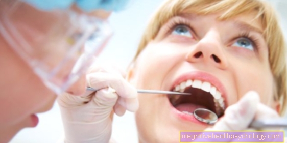
Gums
General
The gums (lat. Gingiva, Greek ulis) is part of the tooth support system and represents the epithelial Part.
Since the gums have a subcutaneous tissue (Subcutis) is missing, it cannot be moved. In addition, the gums cannot be reproduced.

Structure of the gums
Histologically it is Gums from a multilayered squamous epithelium with hardly any horny layers.
Even if the gums cannot be completely reproduced, the mucous membrane has a high regenerative capacity and therefore heals very quickly.
Between everyone tooth and the Gums there is a small gum furrow (Gingival sulcus). If the gums are healthy, this is the furrow approx. 2mm deep. The inner fringing epithelium faces this furrow.
It is divided into the sulcus epithelium, which glides freely on the tooth, and the adhesive epithelium. The Adhesive epithelium is through small connecting cells (Hemidesmosomes) connected to the root cement. The gums are triangular between the individual teeth.
This gum is called the Interdental papilla (Interdental papilla) designated. The borderline between the gums and the dark red oral mucosa, which is movable, is called Mucogingival line (mucogingival border) designated.
clinic
Since the gums can quickly become inflamed, especially in the small furrows (Inflammation of the gums/ Gingivitis), is a regular and thorough cleaning of the Sulci necessary.
This cleaning is a little more difficult because the furrows with the toothbrush difficult to reach. Inflammation of the gums goes with it Toothache, Reddening of the gums and bleeding of the gums. Therapy and prophylaxis are intensive Oral hygiene (dental care). In addition, the gum furrows and depressions can be enlarged and are then referred to as gingival pockets. Periodic pockets of more than 2mm Depth as a disease.


.jpg)